- Cart 0
- English
Caspase-dependent apoptosis at a glance
June 26, 2025
Clicks:430
About the Caspase Family:
The Caspase family is a group of proteases present in the cytoplasm that play important roles in programmed cell death (e.g., apoptosis) and inflammatory processes. Caspase is an abbreviation for "Cysteine-dependent Aspartate-directed proteASEs", which contain cysteine at the active site and can specifically cleave the peptide bond after the aspartate residue of the target protein.
Caspase family
Caspase Family Members:
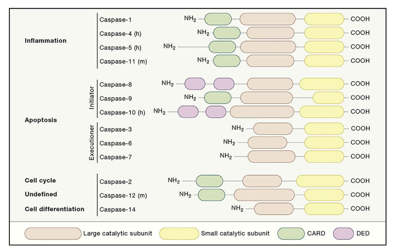
Caspases can be divided into inflammatory caspases and apoptotic caspases according to their function:
Inflammatory caspase: including caspase-1, -4, -5, and -11, these enzymes are mainly involved in the activation of interleukin precursors in the inflammatory response, but are not directly involved in the transduction of apoptosis signals;
Apoptotic caspase: mainly involved in apoptosis, it can be divided into initiating caspase, such as caspase-2, -8, -9, -10, which is responsible for cutting the precursor of effector caspase to produce effector caspase.
Effect caspases: caspase-3, -6, -7, responsible for cleaving structural proteins and regulatory proteins in the nucleus and cytoplasm to inactivate or activate.
Features of apoptosis-related caspase:
Activation mechanism
Self-shearing activation: Many caspase exist in inactive precursor forms that require specific stimuli or interactions with other molecules for self-shearing for activation. For example, caspase-8 and caspase-9 are activated when they bind to specific adaptor proteins to form complexes.
Cascade: Once the initiating caspase is activated, it further activates more effector caspases, forming a cascade that amplifies the signal. This mechanism ensures that apoptosis is an irreversible process.
Substrate specificity
Caspase is highly specific to substrates and typically recognizes aspartate residues in the tetrapeptide sequence and cleavages at this location. Different caspase prefers different cleavage sites, which determine their specific function. Effector caspase can cleave a variety of intracellular proteins, such as lamin, poly-ADP ribose polymerase (PARP), etc., resulting in characteristic changes such as cytoskeletal destruction, DNA fragmentation, and cell membrane alteration.
Structural features
All caspase contains a large catalytic subunit and a small regulatory subunit, which together constitute a heterodimer. Each subunit is composed of multiple functional domains, such as CAPASE Recruitment Domain (CARD) and Death Domain (DD), which are involved in the activation of caspase and its interaction with other proteins.
Regulatory mechanisms
Under normal conditions, caspase is present in the precursor form of the cell and is tightly regulated. Only when the cell receives a specific death signal will it be uninhibited, allowing caspase activation. Some inhibitors such as IAPs (Inhibitor of Apoptosis Proteins) can inhibit apoptosis by directly binding to caspase to block its activity.
Activation of Caspase
The activation process of caspase can be divided into homotypic activation and heterotypic activation, which play a crucial role in the process of apoptosis. The two activation methods and their characteristics are explained in detail below.

Activation of Caspase
1. Homotypic activation
Homologous activation refers to the process by which caspase molecules interact to form dimers or multimers, thereby achieving self-cleavage and activation. This process usually involves the interaction between caspase precursors (procaspase). In homologous activation, an unactivated procaspase interacts with another procaspase through its specific domain, such as CARD or DD, to form a homodimer or multimeric complex. This polymerization state alters the conformation of Procaspase, allowing it to self-shear, removing inhibitory regions, exposing active sites, and ultimately forming a catalytically active mature Caspase.
For example, Caspase-9: In the mitochondrial pathway, cytochrome c is released from the mitochondria to the cytoplasm and combines with Apaf-1 to form apoptosomes. This complex recruits multiple procaspase-9 molecules, prompting their homologous activation;
Caspase-8: In the exogenous pathway, procaspase-8 undergoes homologous activation within DISC through interactions when the death receptor binds to its ligand and recruits adaptor proteins to form DISCs (Death-Inducing Signaling Complex).
2. Heterotypic activation
Heterologous activation refers to the interaction between different types of proteins or members of the Caspase family to promote the activation of each other. This type of activation usually requires the involvement of other cofactors or adapter proteins. In heterologous activation, one procaspase interacts with other non-caspase proteins that may act as activators or scaffolds to aid procaspase localization and conformational alterations. For example, certain adaptor proteins can promote aggregation and conformational changes in procaspase by providing specific binding interfaces, thereby facilitating their self-cleavage and activation.
Such as caspase-3 and caspase-7: although they are primarily activated directly by the initiating caspase as effector caspases, they sometimes require the help of specific scaffold proteins or other regulatory molecules to complete full activation;
Bid protein: In the endogenous pathway, caspase-8 can not only directly activate downstream effector caspase, but also cleave Bid protein to generate tBid, which migrates to mitochondria and further amplifies apoptotic signals, indirectly promoting the activation of other caspase.
Homoactivation relies on direct interactions between procaspase to form homodimers or multimers to promote self-cleavage and activation, commonly seen during activation of initiating caspase such as caspase-8 and caspase-9. Heterologous activation involves the interaction of different proteins, some of which act as activators or scaffolds to help procaspase localize, aggregate, and conformational changes, and are commonly found in complex and multi-step apoptotic signaling pathways.
Caspase-mediated apoptosis pathway
Apoptotic pathways
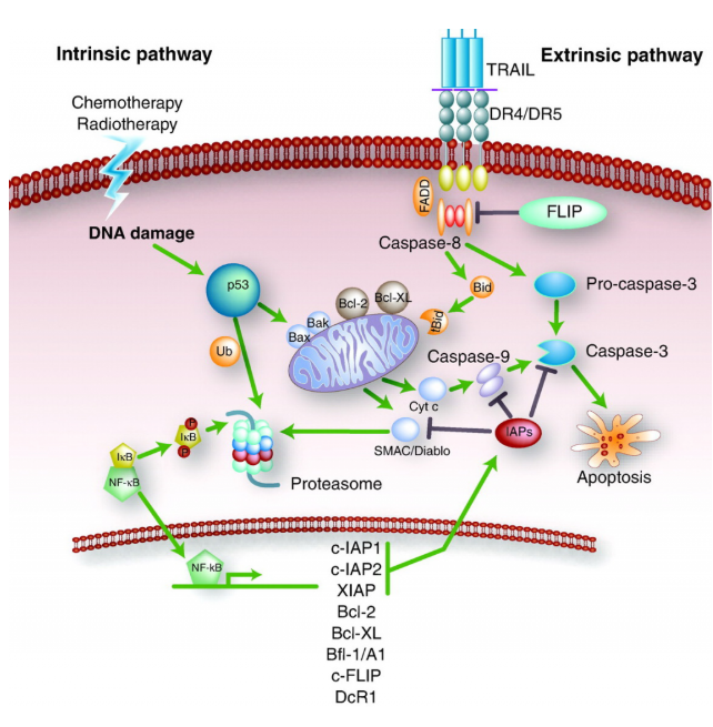
The caspase-mediated apoptosis pathway is an important mechanism of programmed cell death and is mainly divided into two main categories: exogenous (death receptor) pathways and endogenous (mitochondria) pathways. These pathways carry out the process of apoptosis by activating a series of caspase proteases.
1. Exogenous (death receptor) pathway
Initiation: This pathway is activated when specific ligands such as Fas ligands or tumor necrosis factor (TNF) bind to the corresponding receptors on the cell surface. These receptors belong to the TNF receptor family members.
Signaling: When a ligand binds to a receptor, it recruits adaptor proteins and forms a macromolecular complex called a death-inducing signaling complex (DISC) near the cell membrane. This process results in self-cleavage and activation of initiating caspase-8.
Execution phase: activated caspase-8 can directly cleave and activate effector caspases such as caspase-3, -6, -7, and directly trigger the apoptosis program; Alternatively, the mitochondrial pathway is indirectly activated by cleaving the pro-apoptotic protein Bid to further amplify the apoptotic signal.
2. Endogenous (mitochondria) pathways
Triggers: A variety of intracellular stimuli, including but not limited to DNA damage, oxidative stress, hypoxia, cell cycle arrest, etc., may activate this pathway.
Bcl-2 family regulation: Proteins in the Bcl-2 family play a key role in this process. Anti-apoptotic members (e.g., Bcl-2, Bcl-xL) inhibit apoptosis, while pro-apoptotic members (e.g., Bax, Bak) promote apoptosis. When pro-apoptotic signals predominate, Bax/Bak forms pores in the cell membrane, resulting in increased mitochondrial membrane permeability.
Cytochrome C release: With the increase of mitochondrial membrane permeability, cytochrome C is released from the mitochondria into the cytoplasm, where together with other proteins form apoptosomes and activate caspase-9.
Execution phase: Activated caspase-9 then activates downstream effector caspases such as caspase-3, -6, -7, which cleave specific substrates, leading to changes in cellular structure and ultimately the occurrence of apoptosis.
These two pathways, while having different triggering mechanisms, ultimately both perform apoptosis by activating a series of caspases. In addition, it is important to note that in some cases, these two pathways are not completely independent, but can interact and cross-regulate to ensure that the apoptotic process can be carried out accurately and error-free. For example, exogenous pathways can indirectly affect endogenous pathways by activating Bid proteins. Similarly, endogenous pathways can also affect the efficiency and effectiveness of exogenous pathways. This complex network of interactions ensures that apoptosis is realized as a precisely regulated phenomenon of life.
Caspase assays
Caspase detection methods include: flow cytometry, WB, IF, IHC, ELISA, etc., such as:
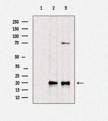
abs132005 Rabbit anti-Cleaved-caspase3 Polyclonal Antibody
Western blot analysis of extracts from different samples was performed using cleaved caspase 3 (Asp175), p17Ab. Lane 1: Rat liver, antigen-specific peptide blocking. The second course: mouse liver. The third course: rat spleen
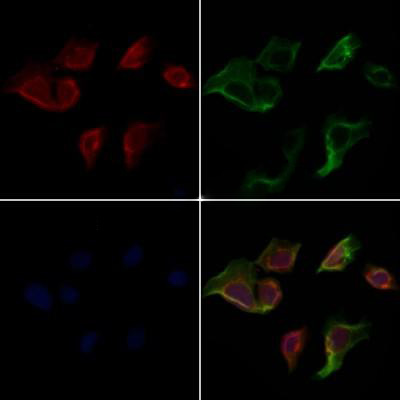
abs132005 Rabbit anti-Cleaved-caspase3 Polyclonal Antibody
HeLa cells were stained by IF/ICC, samples were fixed with PFA, permeabilized in 0.1% Triton X-100, and then blocked in 10% serum for 45 min at 25°C. Samples are then incubated with primary antibody (1:200) and mouse anti-β tubulin antibody (1:200) for 1 h at 37°C. AlexaFluor594-conjugated goat anti-rabbit IgG(H+L)Ab (Red) and AlexaFluor488-conjugated goat anti-mouse IgG(H+L)Ab (Green) were used as secondary antibodies. Nuclear backstaining is DAPI (blue).
Related product recommendations
|
Catalog number |
Product name |
specification |
|
Rabbit anti-Cleaved-caspase3 Polyclonal Antibody |
100ug |
|
|
Rabbit anti-Caspase 3 Polyclonal Antibody |
100uL |
|
|
Rabbit anti-Caspase 9 Polyclonal Antibody |
100uL |
|
|
Mouse anti-Caspase 8 Monoclonal Antibody |
100uL |
|
|
Rabbit anti-Caspase 10 Monoclonal Antibody(2J3 |
100uL |
|
|
Rabbit anti-caspase-9 p10 Polyclonal Antibody |
100uL |
|
|
More CASPASE antibodies can be found on the CASPASE-ABSIN website |
||
Absin provides antibodies, proteins, ELISA kits, cell culture, detection kits, and other research reagents. If you have any product needs, please contact us.
|
Absin Bioscience Inc. |
 Follow us on Facebook: Absin Bio Follow us on Facebook: Absin Bio |