worldwide@absin.cn
  • Sign in or Register
    My account Recent orders Wish list Address management

    Sign out

  • Cart 0
    The latest additions

      0 items in total

      Subtotal $0.00

      Go to cart
    • English
      Chinese
    爱必信(absin)官网
    爱必信(absin)官网

    000000 Citations

    Home
    Products
    Promotions
    Resources
    Product ApplicationFAQsNewsCitationsElectronic DocumentsCalculation Tools
    About us
    Contact us
    worldwide@absin.cn
    • Sign in or Register
      My account Recent orders Wish list Address management

      Sign out

    • Cart 0
      The latest additions

        0 items in total

        Subtotal $0.00

        Go to cart
      • English
        Chinese
      爱必信(absin)官网
      爱必信(absin)官网

      000000 Citations

      Home
      Products
      Promotions
      Resources
      Product ApplicationFAQsNewsCitationsElectronic DocumentsCalculation Tools
      About us
      Contact us
      Home
      abs173744

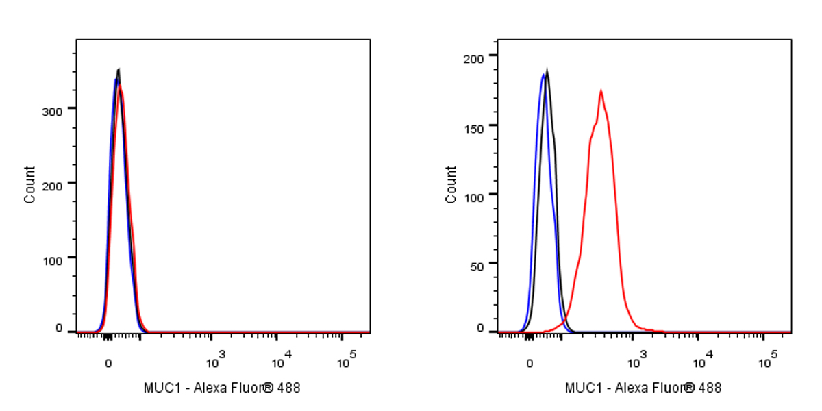
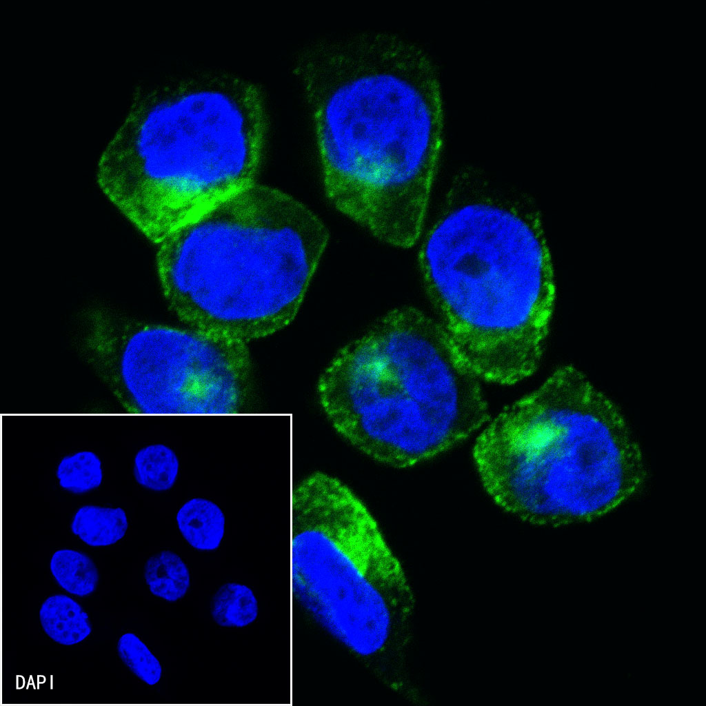
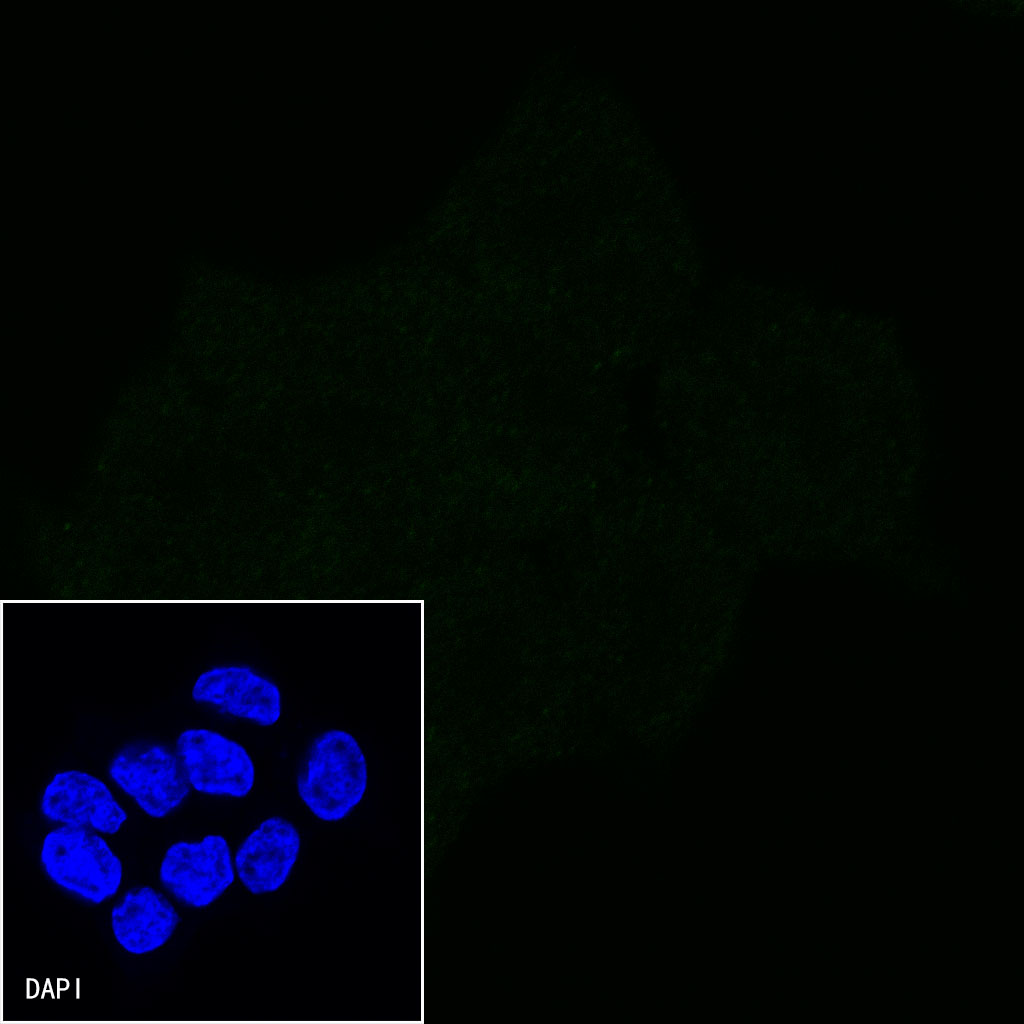
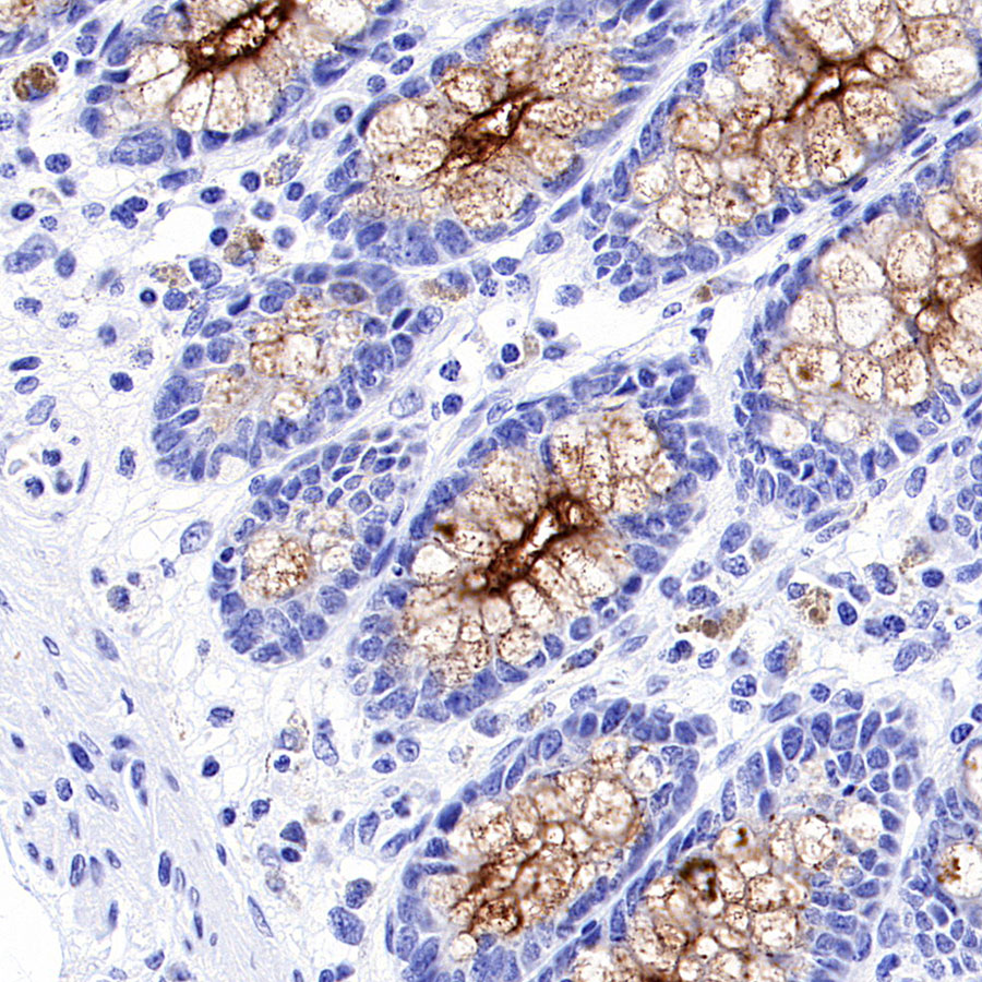
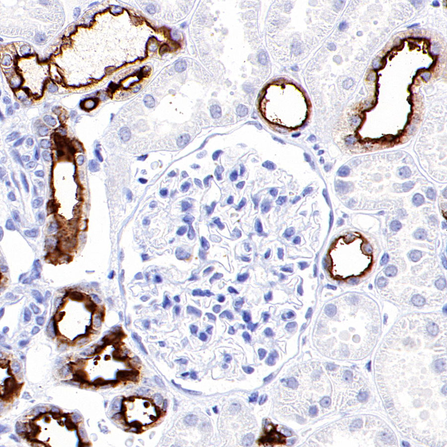
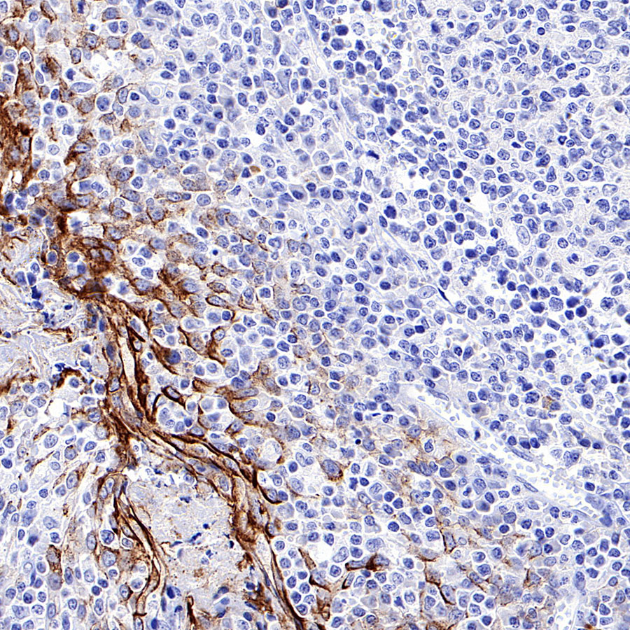
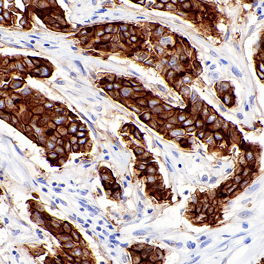
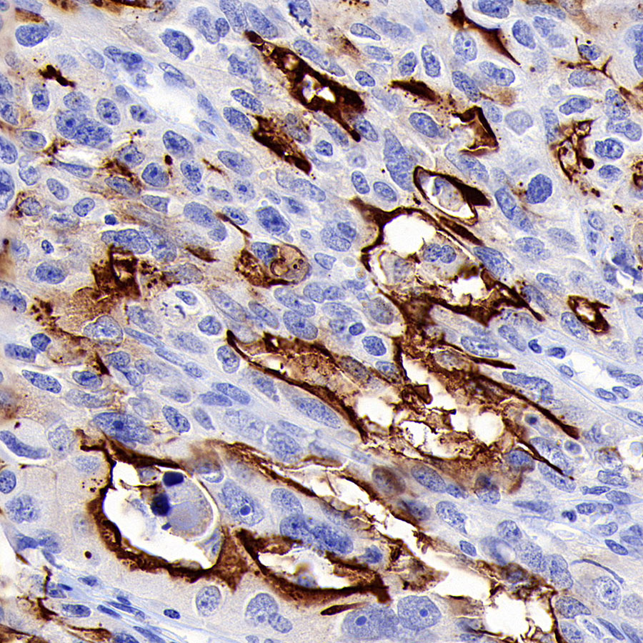
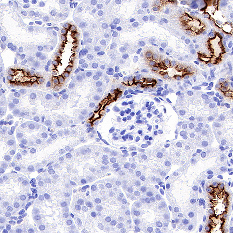
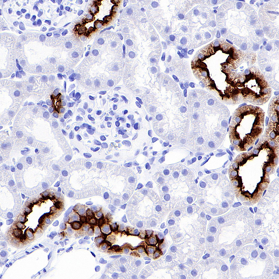
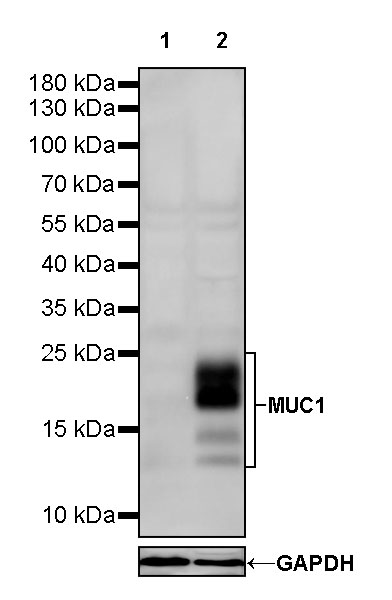
      Mouse anti-MUC1 Monoclonal Antibody(777-71)

      Mucin-1; MUC-1; Breast carcinoma-associated antigen DF3; Cancer antigen 15-3 (CA 15-3); Carcinoma-associated mucin; Episialin; H23AG; Krebs von den Lungen-6 (KL-6); PEMT; Peanut-reactive urinary mucin (PUM); Polymorphic epithelial mucin (PEM); Tumor-associated epithelial membrane antigen (EMA); Tumor-associated mucin,MUC1
      Product ImageProduct ImageProduct ImageProduct ImageProduct ImageProduct ImageProduct ImageProduct ImageProduct ImageProduct ImageProduct Image
      Reactivity:
      Human, Mouse, Rat
      Application:
      WB, IP, IHC-P, IF, ICC, FCM(Intra)
      more>>
      Host:
      Mouse
      Clonality:
      Monoclonal Antibody
      • Datasheet
      • Collect
      • Share:
        Share This on facebook
        Share This on twitter
        Share This on pinterest
        Share This on linkedin
        Share This on skype
        Share This on telegram
        Share This on whatsapp
        Share This on wechat
      Request bulk quotation
      Add to Cart
      • Product Details
      • FAQ
      • Pictures
      • Documents
      Tips : This product is for research use only. Not for use in diagnostic prodcedures.
      Dear customer, if you have any questions or want to know about our products, you can click the "I want to ask" button to fill in your questions. Unsuccessful submission? Please contact worldwide@absin.cn.
      Ask questions
      • Publication date
      • Impact factor
      Search

      Product Album

      Experimental result diagram

      Flow cytometric analysis of 4% PFA fixed 90% methanol permeabilized HCT116 (Human colorectal carcinoma epithelial cell, left) / T47D (Human ductal breast epithelial tumor epithelial cell, Right) labelling MUC1 antibody at 1/10000 dilution (0.01 ug) / (Red) compared with a Mouse monoclonal IgG (Black) isotype control and an unlabelled control (cells without incubation with primary antibody and secondary antibody) (Blue). Goat Anti - Mouse IgG Alexa Fluor® 488 was used as the secondary antibody. Negative control: HCT116

      ICC shows positive staining in T-47D cells. Anti-MUC1 antibody was used at 1/250 dilution (Green) and incubated overnight at 4°C. Goat polyclonal Antibody to Mouse IgG - H&L (Alexa Fluor® 488) was used as secondary antibody at 1/1000 dilution. The cells were fixed with 4% PFA and permeabilized with 0.1% PBS-Triton X-100. Nuclei were counterstained with DAPI (Blue).

      Negative control: ICC shows negative staining in HCT-116 cells. Anti-MUC1 antibody was used at 1/250 dilution and incubated overnight at 4°C. Goat polyclonal Antibody to Mouse IgG - H&L (Alexa Fluor® 488) was used as secondary antibody at 1/1000 dilution. The cells were fixed with 4% PFA and permeabilized with 0.1% PBS-Triton X-100. Nuclei were counterstained with DAPI (Blue).

      IHC shows positive staining in paraffin-embedded human colon. Anti-MUC1 antibody was used at 1/1000 dilution, followed by a HRP Polymer for Mouse & Rabbit IgG (ready to use). Counterstained with hematoxylin. Heat mediated antigen retrieval with Tris/EDTA buffer pH9.0 was performed before commencing with IHC staining protocol.

      IHC shows positive staining in paraffin-embedded human kidney. Anti-MUC1 antibody was used at 1/1000 dilution, followed by a HRP Polymer for Mouse & Rabbit IgG (ready to use). Counterstained with hematoxylin. Heat mediated antigen retrieval with Tris/EDTA buffer pH9.0 was performed before commencing with IHC staining protocol.

      IHC shows positive staining in paraffin-embedded human tonsil. Anti-MUC1 antibody was used at 1/1000 dilution, followed by a HRP Polymer for Mouse & Rabbit IgG (ready to use). Counterstained with hematoxylin. Heat mediated antigen retrieval with Tris/EDTA buffer pH9.0 was performed before commencing with IHC staining protocol.

      IHC shows positive staining in paraffin-embedded human breast cancer. Anti-MUC1 antibody was used at 1/1000 dilution, followed by a HRP Polymer for Mouse & Rabbit IgG (ready to use). Counterstained with hematoxylin. Heat mediated antigen retrieval with Tris/EDTA buffer pH9.0 was performed before commencing with IHC staining protocol.

      IHC shows positive staining in paraffin-embedded human ovarian cancer. Anti-MUC1 antibody was used at 1/1000 dilution, followed by a HRP Polymer for Mouse & Rabbit IgG (ready to use). Counterstained with hematoxylin. Heat mediated antigen retrieval with Tris/EDTA buffer pH9.0 was performed before commencing with IHC staining protocol.

      IHC shows positive staining in paraffin-embedded mouse kidney. Anti-MUC1 antibody was used at 1/1000 dilution, followed by a HRP Polymer for Mouse & Rabbit IgG (ready to use). Counterstained with hematoxylin. Heat mediated antigen retrieval with Tris/EDTA buffer pH9.0 was performed before commencing with IHC staining protocol.

      IHC shows positive staining in paraffin-embedded rat kidney. Anti-MUC1 antibody was used at 1/1000 dilution, followed by a HRP Polymer for Mouse & Rabbit IgG (ready to use). Counterstained with hematoxylin. Heat mediated antigen retrieval with Tris/EDTA buffer pH9.0 was performed before commencing with IHC staining protocol.

      WB result of MUC1 Mouse mAb Primary antibody: MUC1 Mouse mAb at 1/500 dilution Lane 1: HCT-116 whole cell lysate 20 ug Lane 2: T-47D whole cell lysate 20 ug Negative control: HCT-116 whole cell lysate Secondary antibody: Goat Anti-Mouse IgG, (H+L), HRP conjugated at 1/10000 dilution Predicted MW: 122 kDa Observed MW: 24 kDa Exposure time: 2.4 s

      • The batch number you entered does not exist. Please input the correct batch number information
      • Datasheet product specification
      • Products
      • Resources
      • Product Application
      • Calculation Tools
      • FAQs
      • Citations
      • Electronic Documents
      • About us
      • Company Profile
      • Ordering Tools
      • Customer Support
      • Contact us
      Email: worldwide@absin.cn
      picture invalid picture invalid
      ICP:16001690 Absin Bioscience Inc.All rights reserved.