- Cart 0
- English
What you should know about markers in WB experiments
July 10, 2025
Clicks:3434
1. What is a protein marker?
A protein marker, also known as a protein molecular weight marker, is a mixture of proteins of known molecular weight used in protein electrophoresis analysis. They typically contain multiple bands of protein of different molecular weights that migrate different distances during electrophoresis depending on the size of the molecular weight and the difference in charge, resulting in an ordered series of bands on the gel.
2. What is the role of Marker in WB?
Molecular weight indexing: Marker contains a series of proteins of known molecular weight, and the molecular weight of the protein to be tested can be accurately determined by comparing it with the banded protein band on the Marker;
Verify electrophoresis and transfer:Marker moves onto the membrane during electrophoresis and transfer to help researchers validate the effects of these steps. By checking the banding distribution of the Marker, you can be sure that no abnormalities have occurred during electrophoresis and transfer;
Determine the protein migration distance: By measuring the migration distance of the Marker, it is possible to estimate the migration distance of the protein to be measured. This is important for further data analysis and interpretation of results;
Calibrated gel concentration:Marker can also be used to assist researchers in selecting the appropriate gel concentration. Markers of different molecular weights will produce different migration patterns at different gel concentrations, so the appropriate gel concentration can be selected according to the migration characteristics of the Marker.
3. What are the classifications of Marker and what are the characteristics of each?
● Natural Protein Marker:
These markers are proteins extracted from natural sources that undergo natural modification processes such as cleavage, disulfide bond formation, glycosylation, or phosphorylation, and maintain their complex 3D structure. Natural protein markers are closer to their natural state in the body, which makes them very useful in drug development and biomedical research. Natural protein markers can provide a more accurate molecular weight estimate because they have not been reconstituted or synthesized, preserving the natural properties of the protein. abs9919 native protein Marker (66-669kDa), after native electrophoresis, can be stained with Coomassie brilliant blue to obtain 4 bands with uniform distribution and similar density, which is not suitable for SDS-Page, because in the presence of SDS, proteins containing multiple subunits will depolymerize to varying degrees.
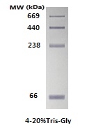
abs9919 native protein Marker (66-669kDa)
● Transgender Marker:
These markers are denatured during the production process, usually by heat and chemicals (e.g., SDS) to ensure that the protein fully unfolds into a linear structure. The advantage of denaturing markers is that they provide consistent protein migration properties, which are important for accurate molecular weight estimation and experimental reproducibility. Due to the denaturation process, the proteins of these markers maintain a consistent charge and shape, making their migration during electrophoresis more predictable.
Denatured protein markers can generally be divided into two types: prestained and non-prestained
● Unstained Protein Marker:
This category of markers includes broad, high, and low molecular weight protein standards. They are the simplest and most accurate of the kind, and because they do not come with a dye molecule or a labeled molecule, the size shown is exactly the original size of the protein. The disadvantage is that it is not visible during electrophoresis, and it needs to be stained after the end of electrophoresis to be visible, and the electrophoresis process cannot be monitored in real time. It is suitable for accurate protein size, but it is relatively cumbersome to perform and requires additional staining steps. For example, abs9840 unstained protein Marker (14.4-97.4kDa), stained with Coomassie Brilliant Blue R-250 can obtain six bands with uniform distribution and same density, the product contains SDS, which is not suitable for non-denaturing gel.
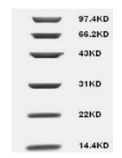
abs9840 Unstained Protein Marker (14.4-97.4 kDa)
● Prestained Protein Marker:
These markers are produced with a dye in bond, so that the bands of different colors can be directly observed during electrophoresis or during transfer. Prestained protein markers allow experimenters to monitor electrophoresis and estimate mobility during and after electrophoresis, as well as after transfer. It is divided into monochrome pre-dyeing and multi-color pre-dyeing. Single-color pre-stained markers often use a doubling of the concentration of some of these bands to indicate their size, while multicolor prestained markers are distinguished by different colors that are easier to identify. It is important to note that the prestained protein marker is covalently coupled to the dye, so there may be some changes in migration characteristics during electrophoresis under different buffer conditions, which may lead to some bias, so it is not suitable for precise protein positioning.

abs923 prestained protein marker (10-245 kDa)
● Pre-stained luminescent Western Marker:
Traditional Western blot assays require the use of a prestained protein marker to track the detection of positive antigen signals and transfer efficiency, but the prestained protein marker cannot be exposed on the film, and the exposure signal needs to be judged based on the prestained protein marker on the membrane. absin's new abs90004 prestained luminescent Western Marker is composed of 9 luminescent proteins and 9 prestained proteins; The luminescent proteins in 9 are 23, 30, 36, 44, 50, 62, 90, 120 and 160KDa, respectively, which can bind to antibodies, so they can expose signals on the same membrane as antigens, and the positive signal can be directly judged by the position of Western Marker; The 9 prestained proteins were 15, 25, 35, 40, 55, 70, 100, 130 and 180 KDa, of which 70 KDa was red and the others were blue, which were visible to the naked eye on the membrane after the transfer was completed.
1) The signal can be detected at the same time as the antigen, which makes the judgment of Western blot results more convenient;
2) The luminescent marker does not couple and label other molecules, and the molecular weight is more accurate;
3) The advantages of combining luminescent markers and prestained markers can not only monitor the transfer but also detect them at the same time as antigens.
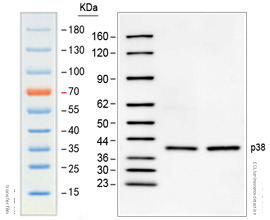
ECL chemiluminescence detection of HRP-conjugated Goat anti mouse IgG secondary antibody (primary antibody: mouse anti P38).
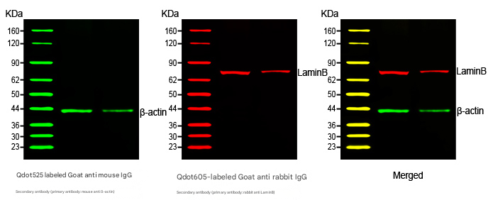
Multiplex fluorescence detection
According to the molecular weight, markers can be divided into high molecular weight, low molecular weight, wide molecular weight, etc. High molecular weight protein markers refer to those labels that are used to detect large molecular weight proteins, and typically contain a high molecular weight range of proteins, and they typically provide molecular weights ranging from tens of kDa to hundreds of kDa. Low-molecular-weight protein markers refer to labels used to detect small-molecular-weight proteins, typically containing proteins with a low molecular weight range, ranging from a few kDa to tens of kDa. Wide-molecular-weight protein markers refer to markers that cover a wide range of molecular weights, often containing proteins with multiple molecular weights ranging from low to high.
4. How to choose a marker? What factors need to be considered?
First of all, look at the type of experiment, non-denatured protein needs non-denaturing protein marker, denatured protein needs denaturing marker, the two can not be replaced by each other, and then look at the size of the target protein, select the corresponding marker, under optional conditions, wide molecular weight protein marker, is a good choice, in addition to the experimental requirements, if you need to detect during electrophoresis and transfer, you need to choose a pre-stained marker, if you need to accurately band molecular weight size, In addition to the above factors, you also need to consider the use system of marker, and the common marker information of ABSIN is as follows:
|
type |
Native protein marker |
Non-prestained protein Marker |
Blue prestained protein Marker |
Multicolor prestained protein Marker |
Prestained luminescent Western Marker |
|||
|
Catalog number |
||||||||
|
Separation volume range |
66-669kDa |
14.4-97.4kDa |
3.3-31kDa |
14.4-97.4kDa |
10-180kDa |
10-245kDa(Tris-glycine-SDS) |
10-180kDa |
Luminesin: 23-160 kDa Prestained protein: 15-180 kDa |
|
Number of strips |
4 |
6 |
5 |
6 |
10 (10 and 17 may not run away due to glue concentration) |
12 |
10 (10 and 17 may not run away due to glue concentration) |
9 |
|
Molecular weight of the band |
66、238、440、669 |
14.4、22、31、43、66.2、97.4 |
3.3、6.5、14.4、20.1、31 |
14.4、20.1、31、43、66.2、97.4 |
10, 17, 25, 33, 43, 55,75, 95, 130,180 |
11、17、20、25、35、48、63、75、100、135、180、135、180、245(Tris-glycine-SDS) |
10, 17, 25, 33, 43, 55,75, 95, 130,180 |
Luminesin: 23, 30, 36, 44, 50, 62, 90, 120, 160 Prestained proteins: 15, 25, 35, 40, 55, 70, 100, 130, 180 |
|
color |
monochrome |
monochrome |
blue |
blue |
75 red, 25k green, the rest blue |
25 green, 75 orange-red, the rest blue |
72 orange, 10 green, the rest blue, 10k bands dark green at 15% glue concentration |
Prestained protein 70KDa is red, others are blue, |
|
Gel systems are recommended |
Native electrophoresis |
Denaturing electrophoresis |
Tricine SDS-PAGE |
There was no change in the apparent molecular weight of Tris-Glycine, Bis-Tris, Tris-Acetate, blue and green bands, and the orange bands were consistent with the inlet |
There was no change in the apparent molecular weight of Tris-Glycine, Bis-Tris, Tris-Acetate, blue and green bands, and the orange bands were consistent with the inlet |
Tris-Glycine |
There was no change in the apparent molecular weight of Tris-Glycine, Bis-Tris, Tris-Acetate, blue and green bands, and the orange bands were consistent with the inlet |
Tris-Glycine, Bis-Tris, etc |
|
dispose |
Coomassie Brilliant Blue staining is required and cannot be heated |
Coomassie brilliant blue staining and a boiling water bath prior to sample loading are required |
No dyeing is required, do not heat |
No dyeing, no heating |
No need to dye, use directly, do not boil, dilute and add reducing agent treatment |
No dyeing, no heating |
No need to dye, use directly, do not boil, dilute and add reducing agent treatment |
Do not dye and use directly, do not boil |
Recommended for non-denaturing protein electrophoresis
|
Catalog number |
Product name |
specification |
|
Lysates for western blotting and immunoprecipitation (WB/IP). |
100mL |
|
|
Tris-Glycine Running Buffer (10×) |
500mL |
|
|
Glass gel Tris-Gly 15%, 10wells, 1.5mm |
10 tablets/box |
|
|
Native Non-Reducing Protein Loading Buffer (5×) |
1mL×5 |
|
|
Native Protein Marker (66-669kDa) |
200uL |
|
|
Coomassie Brilliant Blue Staining Kit (Regular) |
1kit |
WB good things recommended
|
|
Catalog number |
Product name |
specification |
|
Sample processing |
Cytoplasmic, nucleus, and membrane protein extraction kits |
50T |
|
|
Lysates for western blotting and immunoprecipitation (WB/IP). |
100mL |
||
|
Broad spectrum phosphatase inhibitor cocktail (100× stock) |
1mL |
||
|
Broad Spectrum Protease Inhibitor Cocktail (EDTA-free, 100×DMSO stock) |
1mL |
||
|
Protein quantification |
BCA Protein Assay Kit |
500T |
|
|
glue |
Dispensing kits, all kinds of precast gels (refer to the previous table) |
||
|
Loading and electrophoresis |
Loading buffer (2×) |
10mL |
|
|
Electrophoresis Solution(10×) |
100mL |
||
|
Prestained protein marker (10-245 kDa) |
500uL |
||
|
Transfer |
PVDF film (0.45 μm) |
20 sheets |
|
|
NC membrane (0.45 μm) |
20 sheets |
||
|
Electroporation (10×) |
100mL |
||
|
Closed |
Skim milk powder |
500g |
|
|
Bovine serum albumin |
100g |
||
|
Antibody dilution and removal |
Primary and secondary antibody dilutions for WB |
100mL |
|
|
Western primary and secondary antibody removal solution (strongly alkaline) |
500mL |
||
|
Color development and detection |
ECL Chemiluminescence Detection Kit |
25mL×2 |
|
Absin provides antibodies, proteins, ELISA kits, cell culture, detection kits, and other research reagents. If you have any product needs, please contact us.
|
Absin Bioscience Inc. Email: worldwide@absin.net |
 Follow us on Facebook: Absin Bio Follow us on Facebook: Absin Bio |