

- Cart 0
- English
Literature Review: CD28-Targeted Enzyme-Responsive Conformation-Switching Peptide Self-Assembly for Selective T-Cell Acute Lymphoblastic Leukemia (T-ALL) Therapy
October 22, 2026
Clicks:61


Literature Review: CD28-Targeted Enzyme-Responsive Conformation-Switching Peptide Self-Assembly for Selective T-Cell Acute Lymphoblastic Leukemia (T-ALL) Therapy
I. Literature Overview
(1) Basic Information
- Article title: CD28-Targeted Enzyme-Responsive Conformation-Switching Peptide Self-Assembly for Selective T-Cell Acute Lymphoblastic Leukemia (T-ALL) Therapy
- Journal: Advanced Science (a top-tier multidisciplinary journal from Wiley; 2026 IF = 14.1; CAS Q1, TOP)
- DOI: https://doi.org/10.1002/advs.202520963
- Published online: accepted and published in March 2026
(2) Core Conclusions
- Novel target: CD28 is proposed for the first time as a highly specific therapeutic target for T-ALL, circumventing the off-target toxicity of CD3/CD5/CD7-targeted agents that kill normal T cells. T-ALL cells aberrantly overexpress CD28, whereas normal T cells show exhausted CD28 expression in the tumor microenvironment, creating a natural therapeutic window.
- Peptide design: A modular phosphorylated peptide, SAp-CD28, with a three-functional architecture: the CD28-targeting sequence SPMLVAYD, a D-amino-acid phosphorylated self-assembly motif GDFDFDpY, and an NBD fluorescent tracer; excellent water solubility for intravenous administration.
- Enzyme-triggered conformational switching: Tumor-overexpressing alkaline phosphatase (ALP) mediates dephosphorylation, driving an α-helix → β-sheet/β-turn rearrangement that increases CD28 binding affinity 36-fold and in situ forms nano-oligomeric fibers.
- Triple cytotoxic pathways: 鈶?Inhibition of the PI3K–Akt survival pathway: total Akt downregulated >54%, inducing oxidative stress and ROS accumulation; 鈶?Blockade of the PLCγ calcium-signal pathway: total PLCγ decreased 48% and phospho-PLCγ ratio fell 80%, causing intracellular calcium overload, calpain activation, and cytoskeletal collapse; 鈶?Nuclear physical disruption by nanofibers: after endocytic escape, peptides enter the nucleus and continuously assemble long fibers that tear the nuclear membrane, inducing necrotic cell death.
- In vivo efficacy: In a Jurkat xenograft model, single-agent SAp-CD28 achieved a 78.09% tumor inhibition rate; combined with cytarabine, inhibition reached 92.9% with near-complete tumor regression; no pathological damage to major organs, indicating excellent biosafety.
- Generalizability: This enzyme-responsive conformation-switching peptide strategy can be extended to other CD28-overexpressing hematologic malignancies.
(3) Absin Products Used in This Study
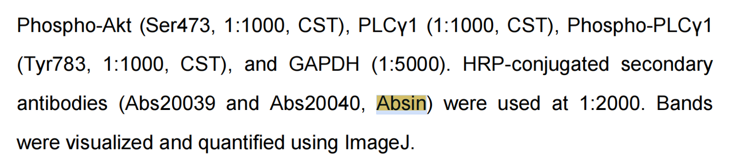
The key Western Blot pathway-validation experiments in this study relied throughout on Absin HRP-conjugated secondary antibodies abs20039 and abs20040, which are core reagents for dissecting the Akt and PLCγ phosphorylation signaling pathways:
- Specifically recognize rabbit monoclonal primary antibodies from CST (total Akt, p-Akt, total PLCγ1, p-PLCγ1), binding precisely to the antibody constant region;
- Conjugated to horseradish peroxidase (HRP) for chemiluminescent imaging that clearly resolves protein bands (corresponding to the original Fig. 5A WB panels);
- High signal-to-noise ratio and extremely low non-specific background enable quantitative visualization of total versus phosphorylated proteins, directly demonstrating SAp-CD28 inhibition of these two key CD28-downstream pathways;
- Supporting grayscale densitometry for statistical analysis, providing gold-standard protein-level validation for transcriptomic differential genes.
II. Research Field Background and Scientific Entry Point
(1) Current State of the Field
- Clinical pain points of T-ALL: T-cell acute lymphoblastic leukemia (T-ALL) is a highly aggressive hematologic malignancy, accounting for ~15% of pediatric and ~25% of adult ALL; adult patients show high relapse, chemoresistance, and extremely poor prognosis. Existing targeted approaches have inherent flaws:
- CD3-targeting clears normal T cells simultaneously, causing fratricide of the T-cell compartment;
- CD5/CD7-targeting exerts strong toxicity against normal immune cells, leading to severe immunosuppression;
- There is a lack of a safe target specifically overexpressed on leukemic cells but low on normal T cells — a brand-new targeting system is urgently needed.
- Gap in CD28 targeting: CD28 is a core costimulatory receptor for T-cell activation; current immunotherapy mostly targets PD-1/CTLA-4, with little development of CD28-targeted agents. Clinical samples confirm aberrant CD28 overexpression on T-ALL cells and CD28 exhaustion in normal T cells within the tumor microenvironment, providing a natural targeting window — yet no CD28-specific nano-peptide drug has been reported.
- Status of enzyme-triggered in situ self-assembly peptides: Enzyme-directed intracellular peptide self-assembly is a recent nanomedicine hotspot that exploits tumor-specific enzymes to trigger in situ nanostructure formation and improve selective killing; however, previous peptides lacked a tumor-specific receptor-targeting module, limiting in vivo enrichment in T-ALL.
- Twofold limitation motivating this study: existing T-ALL drugs have high off-target toxicity, CD28 remains unexploited by peptide nanomedicine, and enzyme-self-assembling peptides lack tumor-specific targeting. This work simultaneously addresses target specificity, enzyme-responsive peptide assembly, and multi-mechanism cytotoxicity, constructing a CD28-targeted enzyme-conformation-switch peptide nano-therapeutic strategy.
(2) Innovative Entry Points of This Study
- Identifies CD28, a T-ALL-specific target with a natural therapeutic window, avoiding damage to normal T cells;
- Engineers a modular phosphorylated peptide that converts from soluble to fiber-forming solely in response to tumor ALP;
- Integrates three killing mechanisms — survival-pathway inhibition, calcium-overload cytoskeletal collapse, and physical nuclear disruption — for multi-layered cytotoxicity;
- Validates pathway inhibition at the protein level with Absin HRP secondary antibodies, anchoring the core mechanistic narrative.
III. Overall Research Logic
The study follows a complete closed loop: target mining → modular molecular design → in vitro physicochemical characterization → cellular targeting/toxicity/mechanism validation → transcriptomic high-throughput pathway screening → protein WB pathway confirmation (supported by Absin secondary antibodies) → in vivo xenograft efficacy and safety evaluation → combination chemotherapy enhancement:
- Target screening: literature and flow cytometry confirmed Jurkat (T-ALL) high CD28 and high ALP, with low expression on normal CD3+ T cells and hepatocytes, locking CD28 as the specific target;
- Peptide design and controls: main molecule SAp-CD28, plus three deletion mutants (SA-CD28p, SAp, CD28p) and a scrambled peptide SAp-CD28-Scr to exclude single-component interference;
- In vitro physicochemical characterization: TEM, FTIR, circular dichroism (CD), and MST affinity assays demonstrated ALP-induced dephosphorylation → α → β conformational switching and greatly enhanced CD28 binding;
- Cellular functional validation: confocal uptake, endocytic route, lysosomal escape, nuclear damage; MTT specificity toxicity; Annexin V flow to define necrosis-dominant death; calcium fluorescence and phalloidin staining for calcium overload and cytoskeletal disruption;
- Omics-driven mechanism screening: RNA-seq differential genes plus KEGG enrichment identified the PI3K–Akt and PLCγ calcium-signaling pathways as core dysregulated routes;
- Protein-level pathway validation (core Absin reagent application): Western Blot of total/phospho Akt and PLCγ1 quantitatively confirmed pathway inhibition, explaining necrosis at the protein level;
- In vivo efficacy: Jurkat nude-mouse xenografts, single-agent and cytarabine-combination dosing, monitoring tumor volume, body weight, serum oxidative marker 8-OHdG, tumor Ki67 proliferation, and organ pathology;
- Mechanism integration and strategy extension: combining signaling-pathway dysregulation with intracellular nanofiber physical nuclear rupture as a dual killing mechanism, proposing a generalizable CD28-targeted enzyme-responsive peptide design paradigm.
IV. Module-by-Module Details and Results
Module 1: Modular peptide design and in vitro physicochemical conformational characterization
Approach: Constructed the tri-functional peptide SAp-CD28 and three controls to dissect the roles of the phosphorylation site, targeting sequence, and assembly motif; simulated the tumor microenvironment with ALP treatment and assessed solubility, assembly morphology, secondary structure, and CD28 binding affinity by multiple techniques.
Results:
- Before enzymatic cleavage all peptides were water-soluble with no Tyndall effect; only SAp-CD28 formed a stable nanofiber gel upon ALP treatment;
- TEM: SAp-CD28 formed 4.15 nm interwoven nanofibers after enzymatic cleavage;
- FTIR + CD: after ALP treatment α-helix content fell 96%, β-sheet rose to 80.3% and β-turn to 17.9%;
- MST affinity: after cleavage SAp-CD28 KD dropped from 293.8 μM to 8.1 μM (36-fold increase), whereas control peptides showed no improvement.
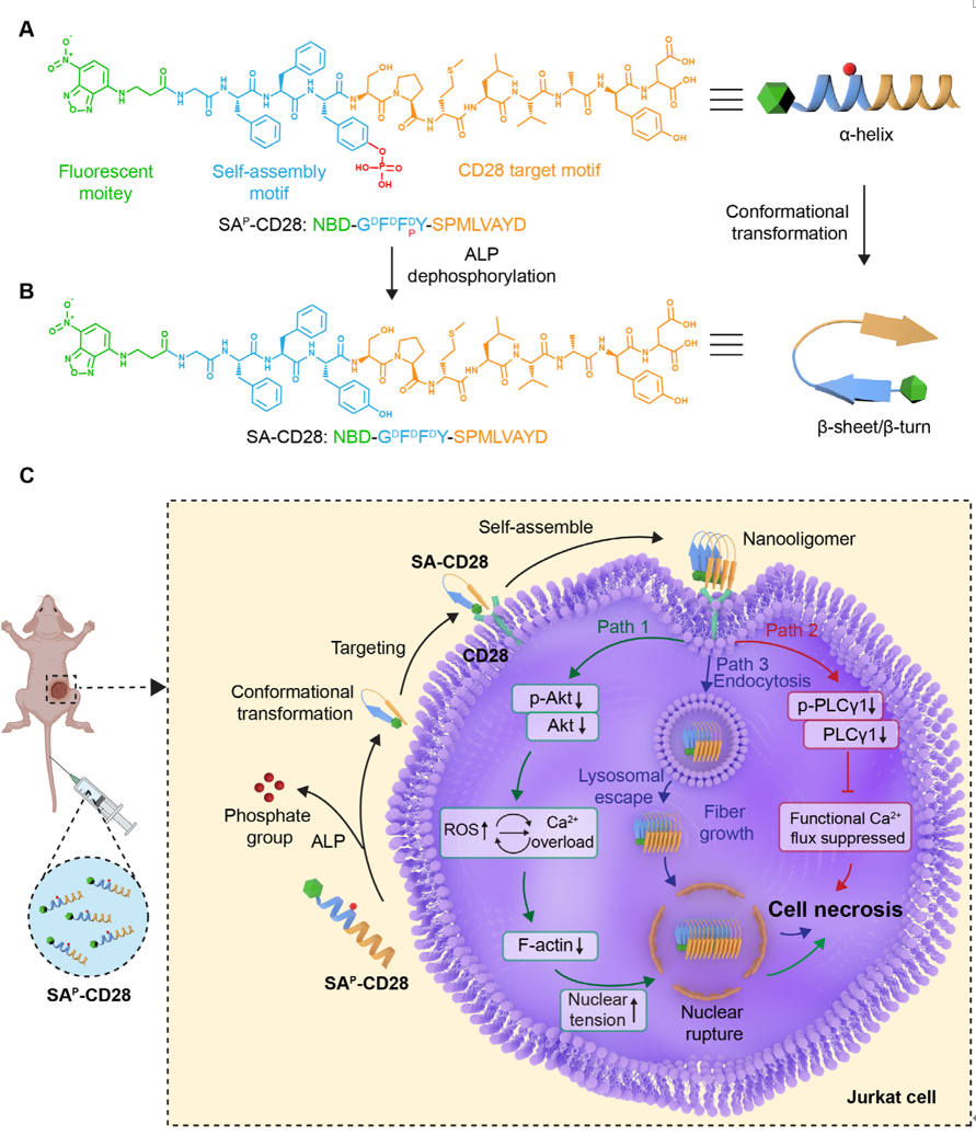
Figure 1. Schematic of peptide molecular design and its tumor-killing mechanism. (A) Chemical structure and secondary conformation of SAp-CD28. (B) Chemical structure and secondary conformation of SA-CD28, generated via alkaline phosphatase-mediated dephosphorylation of SAp-CD28. (C) Schematic illustration of SAp-CD28 inducing nuclear rupture through dysregulation of CD28 downstream signaling and physical damage, and its synergy with the chemotherapeutic agent cytarabine.
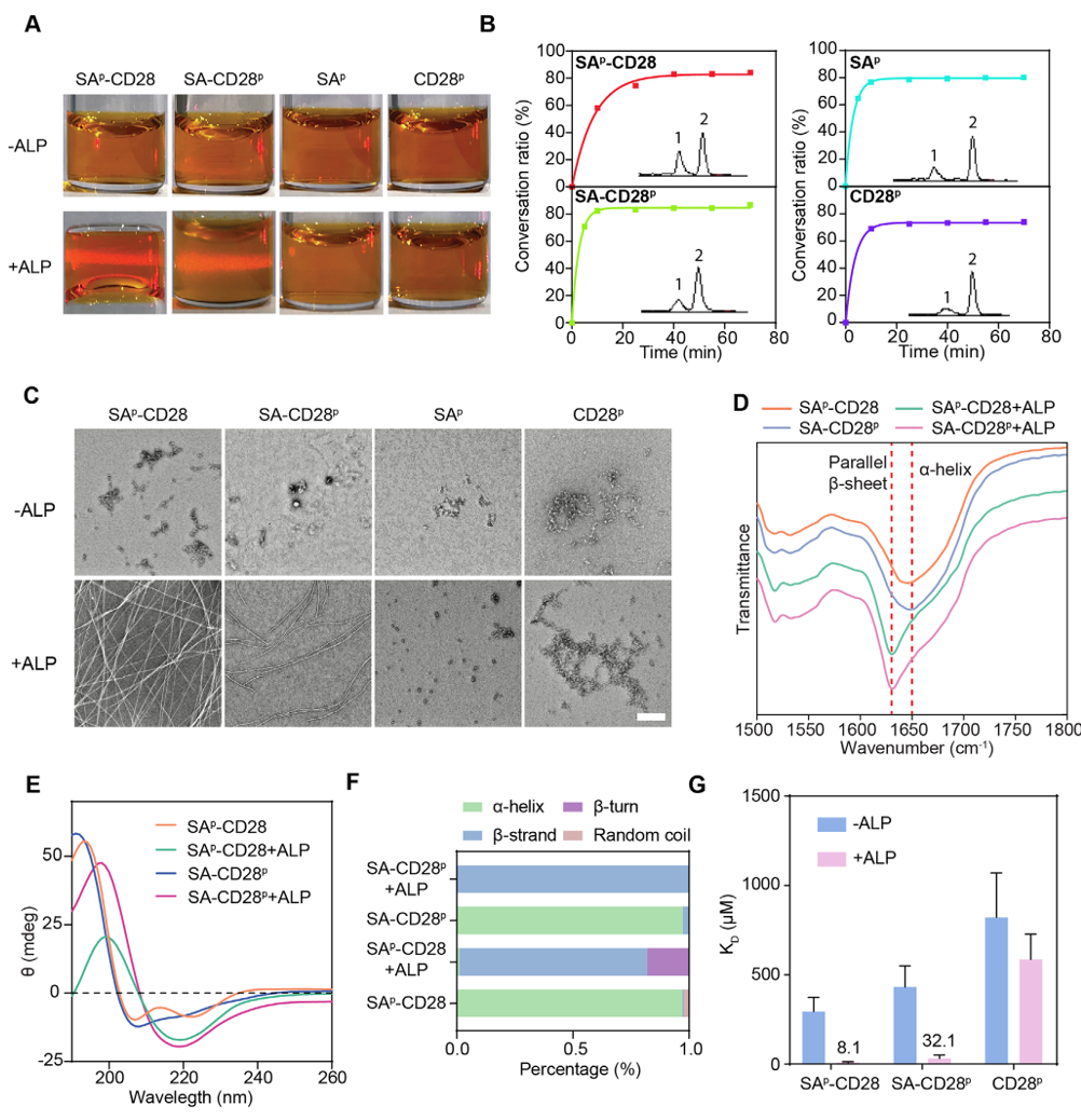
Figure 2. Characterization of the four peptide molecules before and after ALP dephosphorylation. (A) Tyndall effects without or with ALP for 12 h. (B) Conversion rates over time with ALP (10 U/mL); HPLC chromatograms (insets) show peaks before (peak 1) and after (peak 2) dephosphorylation. (C) TEM of the indicated peptides without or with ALP for 12 h (scale bar, 200 nm). (D) FTIR spectra with or without ALP (10 U/mL); dashed lines mark parallel β-sheets and α-helices. (E) CD spectra with or without ALP. (F) Secondary-structure calculation. (G) MST analysis of SAp-CD28, SA-CD28p and CD28p binding to recombinant human CD28 before and after ALP; KD values from 16-point titration (mean ± SD, n = 3).
Module 2: Cellular targeting, uptake, specific cytotoxicity, and subcellular damage
Approach: Using Jurkat T-ALL cells as the model and normal CD3+ T cells and LO2 hepatocytes as safety controls; confocal imaging of uptake and nuclear fragmentation; endocytic inhibitors to define uptake routes; MTT for viability; flow cytometry to distinguish apoptosis/necrosis; Bio-EM to observe nuclear-membrane ultrastructure damage.
Results:
- SAp-CD28 specifically enriched in Jurkat, escaped lysosomes into the nucleus, and caused chromatin condensation and nuclear-membrane rupture; control peptides showed almost no intracellular uptake;
- Uptake depended on microtubule, dynein, and macropinocytosis pathways;
- At 16 μM, Jurkat viability was only 20%, whereas normal T cells and hepatocytes remained >85% viable — excellent selectivity;
- Death was predominantly necrotic (78.03% necrotic cells).
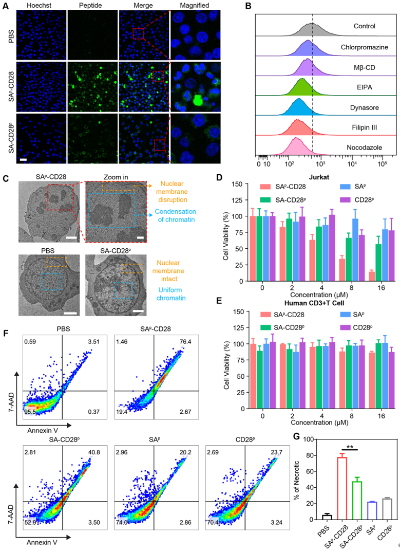
Figure 3. Nuclear damage and apoptosis triggered by peptide self-assembly at the cellular level. (A) CLSM images of Jurkat cells treated with SAp-CD28 or SA-CD28p (scale bar, 25 μm). (B) Flow-chart of fluorescence intensity of Jurkat cells pretreated with internalization inhibitors then incubated with SAp-CD28 for 1 h. (C) Bio-EM imaging after 8 h treatments (scale bar, 500 nm). (D, E) Relative viabilities of (D) Jurkat and (E) human CD3+ T cells with four peptides for 48 h (n = 4). (F) Annexin V-PE/7-AAD apoptosis detection after 12 h. (G) Quantitative apoptosis flow analysis (mean ± SD, n = 3; one-way ANOVA, **p < 0.01).
Module 3: Transcriptomic high-throughput pathway screening
Approach: RNA-seq of PBS-, SA-CD28p-, and SAp-CD28-treated cells; differential-gene screening and KEGG enrichment to locate calcium signaling, PI3K–Akt, and focal-adhesion (cytoskeleton) dysregulated pathways as candidates for WB validation.
Results: SAp-CD28 treatment produced 278 differentially expressed genes, significantly enriched in PI3K–Akt, calcium signaling, and focal-adhesion pathways, indicating survival-pathway inhibition and calcium-homeostasis imbalance.
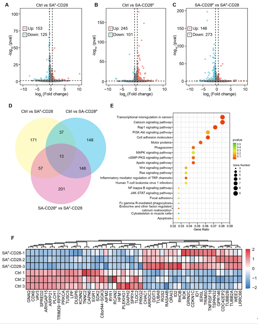
Figure 4. Transcriptomic profiling of peptide-induced apoptosis. (A–C) Volcano plots of gene regulation in Jurkat cells: Control vs SAp-CD28 (A), Control vs SA-CD28p (B), SA-CD28p vs SAp-CD28 (C). (D) Venn diagram of common and unique deregulated genes vs PBS. (E) KEGG pathway enrichment of differential pathways (SAp-CD28 vs PBS). (F) Heatmap of typical apoptosis- and nuclear-damage-associated genes (SAp-CD28 vs PBS).
Module 4: Western Blot protein-pathway validation (core Absin abs20039/abs20040 application module)
Approach: Targeted the transcriptomically enriched Akt and PLCγ pathways; used specific rabbit primary antibodies for total and phosphorylated proteins; incubated with Absin HRP-conjugated secondary antibodies and imaged; grayscale densitometry clarified SAp-CD28 inhibition of the two pro-survival pathways; calcium fluorescence and cytoskeletal staining verified downstream calcium overload and cytoskeleton collapse.
Absin-containing key steps: After electrophoresis and transfer, membranes were blocked overnight; rabbit primary antibodies (Akt, p-Akt, PLCγ1, p-PLCγ1) were incubated; after washing, diluted Absin abs20039/abs20040 HRP-conjugated goat-anti-rabbit secondary antibodies were incubated 1 h at room temperature; thorough TBST washing; HRP chemiluminescent imaging captured bands; ImageJ quantified grayscale values.
Results:
- Total Akt protein downregulated >54%, p-Akt activation markedly reduced — PI3K–Akt survival pathway blocked;
- Total PLCγ1 decreased 48%, phospho-PLCγ/total PLCγ ratio fell 80% — classical calcium-mobilization pathway suppressed;
- Intracellular calcium fluorescence intensity increased 1.4-fold and F-actin cytoskeleton fragmented, confirming calpain-mediated cytoskeletal collapse.
Key value of Absin products: The only immunological detection reagent in this module, it directly enables quantitative visualization of pathway proteins, completes protein-level validation of the transcriptomic data, and underpins the core killing-mechanism derivation.
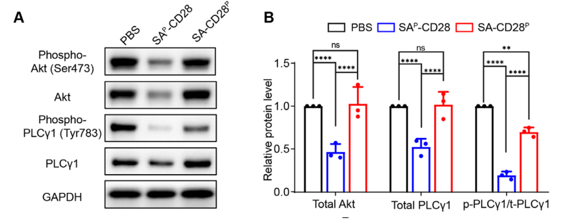
Figure 5. SAp-CD28 disrupts CD28 downstream signaling and induces calcium overload and cytoskeletal collapse. (A, B) Representative Western Blot bands (A) and quantitative analysis (B) of phospho-Akt, Akt, phospho-PLCγ, and PLCγ in Jurkat cells treated with SAp-CD28 or SA-CD28p vs PBS controls.
Module 5: In vivo nude-mouse xenograft efficacy and safety evaluation
Approach: Subcutaneous Jurkat xenografts in nude mice assigned to five groups: PBS, single-agent cytarabine, SA-CD28p, SAp-CD28 single-agent, and SAp-CD28 + cytarabine combination; 28-day dosing, monitoring tumor volume and body weight; endpoint serum oxidative-damage marker 8-OHdG, tumor H&E necrosis staining, Ki67 proliferation staining, and heart/liver/spleen/lung/kidney histopathology.
Results:
- SAp-CD28 single-agent inhibition 78.09%, combination 92.9%, with slight tumor regression;
- No significant body-weight loss or systemic toxicity in any group;
- Combination-group tumor necrosis area >50%, Ki67 proliferation marker dropped to 20%; no tissue lesions in major organs.
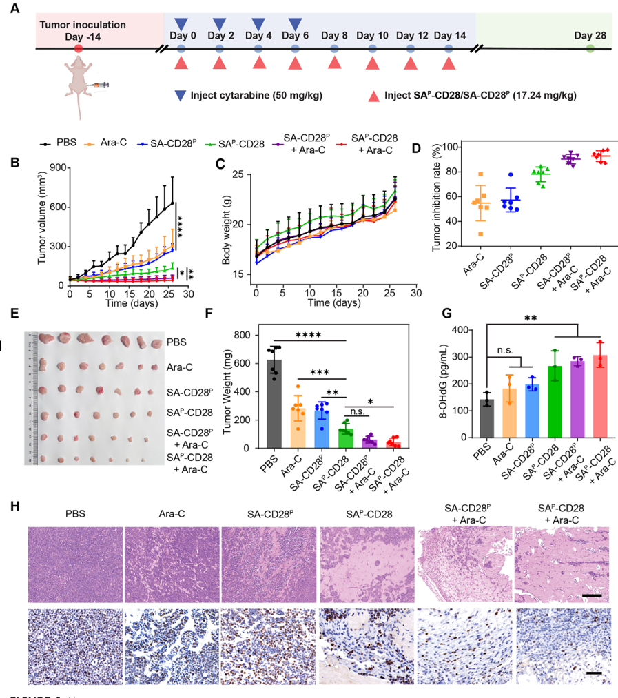
Figure 6. Antitumor effects of the self-assembling peptides in Jurkat tumor-bearing nude mice. (A) Schematic of tumor inoculation and treatment. (B) Tumor growth curves during treatment. (C) Mouse body-weight variations. (D) Tumor inhibition rates at endpoint. (E) Dissected-tumor photographs. (F) Average tumor weight at endpoint. (G) Serum 8-OHdG quantification. (H) Representative H&E (top; scale bar, 200 μm) and Ki-67 IHC (bottom; scale bar, 50 μm) staining (mean ± SD; n = 7 per group for B–D, F; n = 3 for G; two-way or one-way ANOVA; *p < 0.05, **p < 0.01, ***p < 0.001, ****p < 0.0001).
V. Overall Summary of Absin (abs20039, abs20040) Product Role
Throughout this complete CD28-targeted peptide T-ALL therapeutic study, the Absin HRP-conjugated secondary antibodies abs20039 and abs20040 were the irreplaceable core supporting reagents for signaling-pathway mechanism validation:
- Data-reliability guarantee: Compatible with commercial rabbit total/phosphorylated-protein primary antibodies, with strong specificity and few non-specific bands, clean quantifiable WB bands ensured the statistical credibility of the Akt/PLCγ pathway-inhibition conclusions;
- Mechanistic logic linkage: Building on transcriptomic high-throughput screening, they explained at the molecular-protein level the upstream signaling dysregulation (oxidative stress, calcium overload, necrosis) induced by the peptide, constructing the complete mechanistic chain "peptide binds CD28 → pathway inhibition → multi-mechanism cell death";
- Foundation for top-journal publication: Advanced Science demands stringent WB imaging quality and quantitative reproducibility; Absin secondary antibodies give stable luminescence with minimal inter-batch variation, meeting top-journal figure-standardization requirements;
- General research-tool value: These Absin secondary antibodies are widely applicable to tumor signaling-pathway (PI3K/Akt, PLCγ/calcium) WB validation, providing a standardized immunological detection solution for hematologic-malignancy, peptide nanomedicine, and targeted-small-molecule mechanistic research.
Contact Absin
Absin provides antibodies, proteins, ELISA kits, cell culture, detection kits, and other research reagents. If you have any product needs, please contact us.
| Absin Bioscience Inc. worldwide@absin.cn |
 Follow us on Facebook: Absin Bio |