

- Cart 0
- English
Literature Review: TFPI2 Promotes NK Cell–Mediated Glioblastoma Killing through Adhesion and Checkpoint Control
October 22, 2026
Clicks:61


Literature Review: TFPI2 Promotes NK Cell–Mediated Glioblastoma Killing through Adhesion and Checkpoint Control
I. Literature Overview
(1) Basic Information
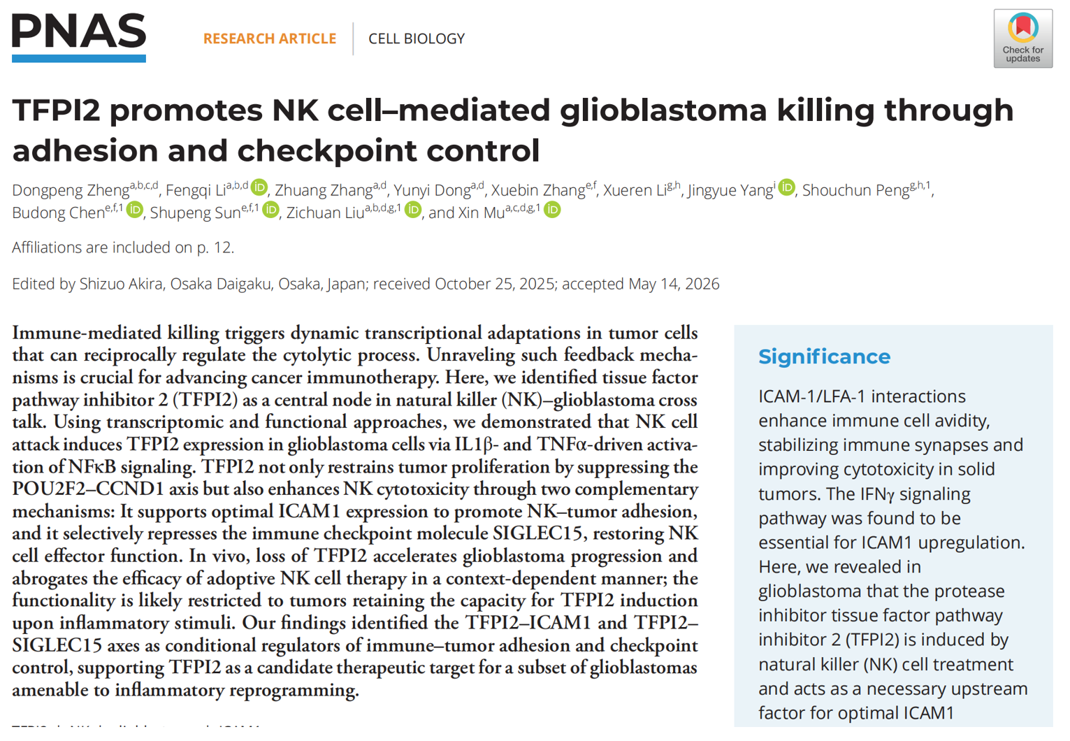
- Article title: TFPI2 promotes NK cell–mediated glioblastoma killing through adhesion and checkpoint control
- Journal: PNAS (Proceedings of the National Academy of Sciences, a leading multidisciplinary journal, 2026 IF ≈ 12.7)
- Volume/issue: 2026, Vol. 123, No. 25, e2529973123
- DOI: https://doi.org/10.1073/pnas.2529973123
- Published online: June 18, 2026
(2) Core Conclusions
- Upstream induction mechanism: Upon contact with glioblastoma (GBM) cells, NK cells secrete IL1β and TNFα, which jointly activate the canonical/non-canonical NF-κB pathways in GBM cells and specifically induce TFPI2 expression. This induction occurs only in GBM cells (U87MG, U138, A172); non-brain tumor cells such as HeLa and A549 do not respond.
- Tumor-cell-autonomous tumor-suppressive pathway: TFPI2 inhibits the transcription factor POU2F2, downregulates cyclin CCND1, and arrests cells in S phase, directly suppressing GBM proliferation and 3D spheroid formation.
- Dual immune-enhancement axes (two core mechanisms):
- TFPI2–LGALS1–ICAM1 adhesion axis: TFPI2 directly binds LGALS1, inhibits its ubiquitin–proteasome degradation, stabilizes LGALS1 protein, and persistently upregulates the adhesion molecule ICAM1, strengthening the NK–tumor immune synapse and enhancing NK cell adhesion, infiltration, and killing;
- TFPI2–SIGLEC15 checkpoint axis: TFPI2 selectively suppresses the immune checkpoint SIGLEC15 (without regulating PD-L1), relieving SIGLEC15-mediated inhibition of NK cells and restoring NK IFNγ secretion and cytotoxic activity.
- Stratified in vivo validation: In the GL261 mouse intracranial model (inflammation-inducible Tfpi2), Tfpi2 knockdown accelerated tumor growth, significantly reduced intracranial NK infiltration, and completely abrogated NK adoptive therapy; depletion of endogenous NK cells produced no additional tumor-promoting effect, confirming that TFPI2 tumor suppression depends entirely on NK cells. In the CT-2A mouse GBM model, IL1β/TNFα stimulation failed to upregulate Tfpi2 and knockdown produced no tumor phenotype, demonstrating pathway heterogeneity. In NCG humanized intracranial xenografts, U87MG-TFPI2 knockout mice infused with human PB-NK cells showed no obvious tumor suppression, validating conservation in the human system.
- Translational significance: TFPI2 may serve as a GBM immune-classification biomarker; elevating tumor TFPI2 expression via STING agonists or demethylating agents, combined with adoptive NK cells and SIGLEC15 blockade, may improve GBM immunotherapy outcomes.
(3) Absin Products Used in This Study
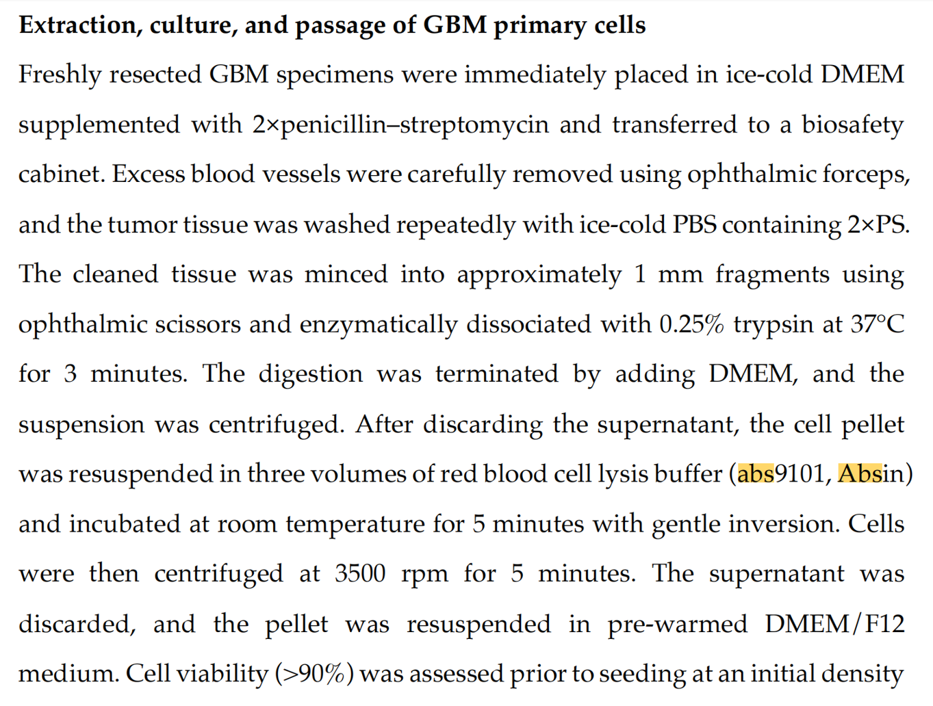
Absin abs9101 Red Blood Cell Lysis Buffer was the key preprocessing reagent for immune cell isolation:
- Human peripheral blood NK preparation: After Ficoll gradient isolation of PBMCs from healthy donor peripheral blood, abs9101 lysed residual red blood cells, removing hemoglobin and erythrocyte debris to ensure PBMC purity for subsequent NK expansion and co-culture;
- Mouse brain tumor-infiltrating lymphocyte (TIL) extraction: Intracranial GL261/CT-2A tumor tissues were enzymatically digested into single-cell suspensions, and abs9101 was used to clear erythrocytes, eliminating flow cytometry background interference and ensuring accurate CD45⁺CD3⁻NK1.1⁺ NK cell gating;
- Sample protection advantage: abs9101 selectively lyses anucleate erythrocytes without damaging nucleated immune cells such as NK and T cells, maintaining stable cell viability and meeting the requirements of flow cytometric staining, in vitro killing, and in vivo adoptive transfer.
II. Research Field Background and Scientific Entry Point
(1) Current State of the Field
- Glioblastoma (GBM) is the most malignant primary brain tumor in adults; standard surgery plus temozolomide chemoradiotherapy yields extremely poor prognosis, with a 5-year survival rate below 10%. The blood–brain barrier and a highly immunosuppressive microenvironment greatly limit the efficacy of CAR-T and immune checkpoint monotherapies.
- NK cell advantages: no MHC-restricted antigen presentation required, low graft-versus-host risk, making them a preferred option for solid tumor adoptive immunotherapy. However, GBM employs multiple immune escape mechanisms that weaken NK cell function: (1) secretion of inhibitory factors such as TGFβ and downregulation of NKG2D ligands; (2) low ICAM1 expression, impairing NK–tumor cell adhesion; (3) high expression of the novel inhibitory checkpoint SIGLEC15, which suppresses NK cytotoxicity independently of the PD-L1 pathway.
- Research gap: previous studies mostly engineered NK cells themselves, with little research on feedback regulatory molecules produced by tumor cells in response to NK attack; TFPI2 function is controversial — it acts as a tumor suppressor in most cancers but can promote stemness in GBM stem cells, and its role in NK–GBM interactions was entirely unknown; no upstream intratumoral regulatory target of SIGLEC15 had been reported.
- Clinical pain point: GBM shows marked immune heterogeneity; some patients do not respond to immunotherapy at all, and stratification biomarkers are lacking.
(2) Innovative Entry Points of This Study
- Novel perspective: focusing on TFPI2, a feedback regulatory molecule produced by GBM cells after NK stimulation, mining the tumor's own bidirectional regulatory circuit (proliferation suppression + immune microenvironment remodeling);
- Mechanistic innovation: simultaneously dissecting three independent pathways — cell cycle, cell adhesion, and a novel immune checkpoint — and establishing the TFPI2–LGALS1–ICAM1 and TFPI2–SIGLEC15 functional axes;
- Heterogeneity explanation: based on multiple human/mouse cell lines and in vivo models, clarifying that only inflammation-sensitive GBM can activate the TFPI2 pathway, providing a basis for patient immune stratification;
- Experimental support: abs9101 enabled purification of human/mouse NK cells and the establishment of standardized in vitro and in vivo immune interaction systems, ensuring reliable immunological data.
III. Overall Research Logic
The study follows a complete closed loop: clinical bioinformatics screening → in vitro induction pathway dissection → cell-autonomous proliferation mechanisms → immunoregulatory molecular interactions → in vitro 2D/3D functional validation → multi-level human/mouse in vivo models → translational inference.
- Clinical cohort mining: TCGA and CGGA human GBM cohorts were stratified into high/low activated-NK-infiltration groups, and the differentially expressed gene TFPI2 was identified; clinical tissue IHC confirmed a positive correlation between TFPI2 and CD56⁺ NK infiltration;
- Upstream pathway analysis: NK-derived IL1β/TNFα activates NF-κB to induce TFPI2, distinguishing direct-contact versus cytokine-mediated induction;
- Proliferation suppression mechanism: TFPI2 knockdown/rescue cells defined the POU2F2–CCND1 cell cycle axis;
- Immune adhesion mechanism: IP-MS identified LGALS1 as a TFPI2-binding protein, and ubiquitination assays elucidated the ICAM1 upregulation mechanism;
- Checkpoint regulation: transcriptomic screening identified SIGLEC15 as a specifically suppressed target of TFPI2, validated by rescue and double-knockdown experiments;
- In vitro function: 2D LDH killing, 3D tumor spheroid co-culture, and NK adhesion assays all relied on abs9101-purified human/mouse NK cells;
- Stratified in vivo validation: three animal models — immunocompetent GL261, immune-cold CT2A, and NCG humanized xenografts — with flow cytometric TIL detection using abs9101-processed single-cell suspensions;
- Translational inference: TFPI2 proposed as a prognostic stratification biomarker and a novel strategy combining inflammation induction with adoptive NK therapy.
IV. Module-by-Module Details and Results
Module 1: Clinical bioinformatics + cell screening identifying TFPI2 as an NK-specifically induced GBM molecule
Approach: TCGA/CGGA bioinformatic screening of differentially expressed genes in high-NK-infiltration groups; NK stimulation validation in human GBM cells, patient-derived primary cells, and control tumor cells (HeLa/A549); clinical tissue IHC correlating TFPI2 with NK infiltration.
Results:
- TFPI2 mRNA was significantly upregulated in the high activated-NK groups of TCGA and CGGA, with Venn intersection locking the candidate molecule (Fig. 1A, SI Fig. S1H);
- Time-gradient stimulation of U87MG/U138/A172 with NK92 showed sustained TFPI2 protein upregulation; cervical cancer and lung cancer cells showed no induction (Fig. 1C-E, SI Fig. S2C-D);
- TFPI2 increased in NK-treated primary GBM cells from 4 patients (Fig. 1G);
- Human GBM tissue IHC: TFPI2 expression was significantly positively correlated with CD56⁺ NK infiltration (Fig. 1H-J).
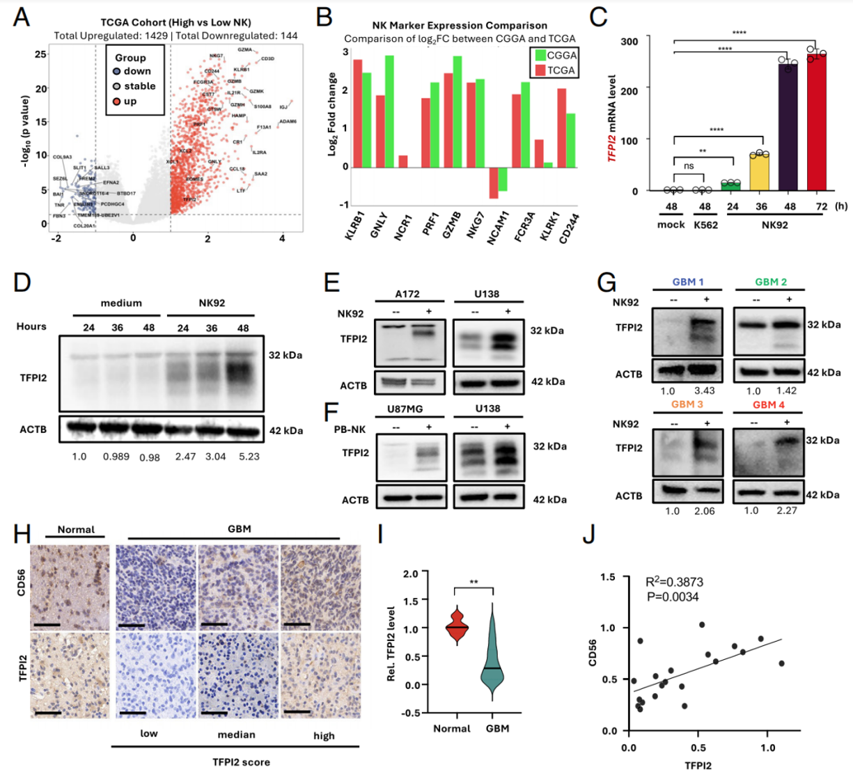
Fig. 1. (A) Volcano plots displaying differentially expressed genes (DEGs) between high-activated (n = 45) and low-activated (n = 53) NK infiltration groups in the TCGA GBM cohort. (B) Log2 fold change of 10 selected NK cell signature genes. (C) U87MG cells cocultured with NK92 cells (E:T ratio = 3:1) or K562 cells for indicated hours; RNA analyzed by RT-qPCR. (D) Western blotting of U87MG cells cocultured with NK92. (E) TFPI2 protein in A172 and U138 GBM cells following NK92 stimulation. (F) TFPI2 protein in U87MG and U138 cells stimulated with PB-NK. (G) TFPI2 protein in primary GBM cells from four patients after NK92 coculturing. (H) CD56⁺ cell infiltration and TFPI2 level in GBM or normal brain tissues (representative IHC; scale bar, 50 μm). (I) Quantification of TFPI2 and CD56 levels. (J) Correlation analysis of CD56 and TFPI2 in GBM (n = 20).
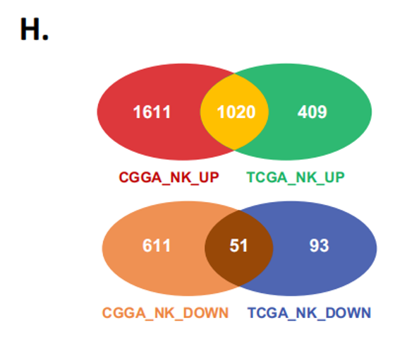
(H) Venn diagram of upregulated and downregulated genes from TCGA and CGGA GBM cohorts.
Module 2: NK induces TFPI2 transcription via the IL1β/TNFα–NF-κB pathway
Approach: Transwell assays to distinguish direct contact from soluble factors; RNA-seq enrichment of the NF-κB pathway; inhibitors, promoter truncation, and RELB binding-site mutation to verify transcriptional regulation; individual and combined cytokine stimulation to confirm upstream signals.
Results:
- Both direct NK contact and supernatant factors induced TFPI2, with stronger effects from direct contact (Fig. 2A);
- NF-κB pathway was significantly enriched in the NK-stimulated GBM transcriptome (Fig. 2B, SI Fig. S3B-D);
- NF-κB inhibitors BMS and BOT64 completely blocked TFPI2 upregulation (Fig. 2C);
- The −254~+100 bp TFPI2 promoter region was the core activation region; mutation of the RELB binding motif abolished inducibility (Fig. 2D-F);
- NK stimulation first induced IL1β, then upregulated TFPI2; IL1B knockdown combined with TNFα neutralization significantly inhibited TFPI2 expression (Fig. 2G-J).
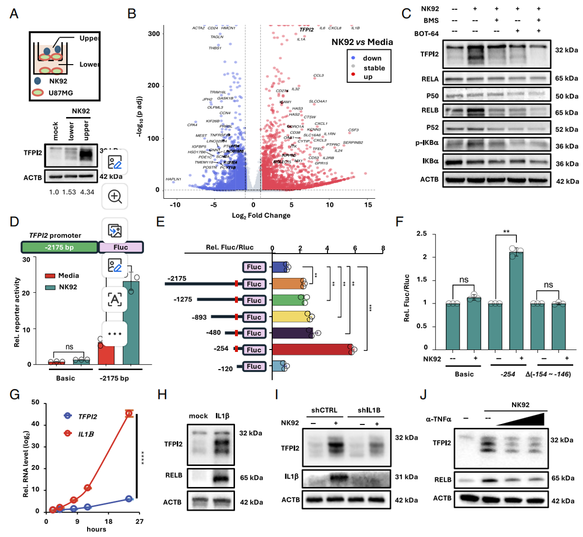
Fig. 2. NK cells induce TFPI2 expression through activation of the NF-κB signaling pathway. (A) TFPI2 protein in upper vs. lower chambers of a Transwell coculture with NK92 cells. (B) Volcano plot of U87MG cells with or without NK92 stimulation. (C) TFPI2 and NF-κB pathway components after BOT64 or BMS-345541 pretreatment and NK92 stimulation. (D) Luciferase activity of pGL3-TFPI2-transfected U87MG cells after NK92 stimulation. (E) Truncated TFPI2 promoter constructs. (F) Luciferase activity of truncated promoters. (G) Time course of IL1β and TFPI2 mRNA induction. (H) TFPI2 and RELB protein after recombinant human IL1β treatment. (I) TFPI2 and IL1β protein in IL1B-knockdown U87MG cells upon NK92 stimulation. (J) TFPI2 induction in the presence of anti-TNFα neutralizing antibody vs. isotype control.
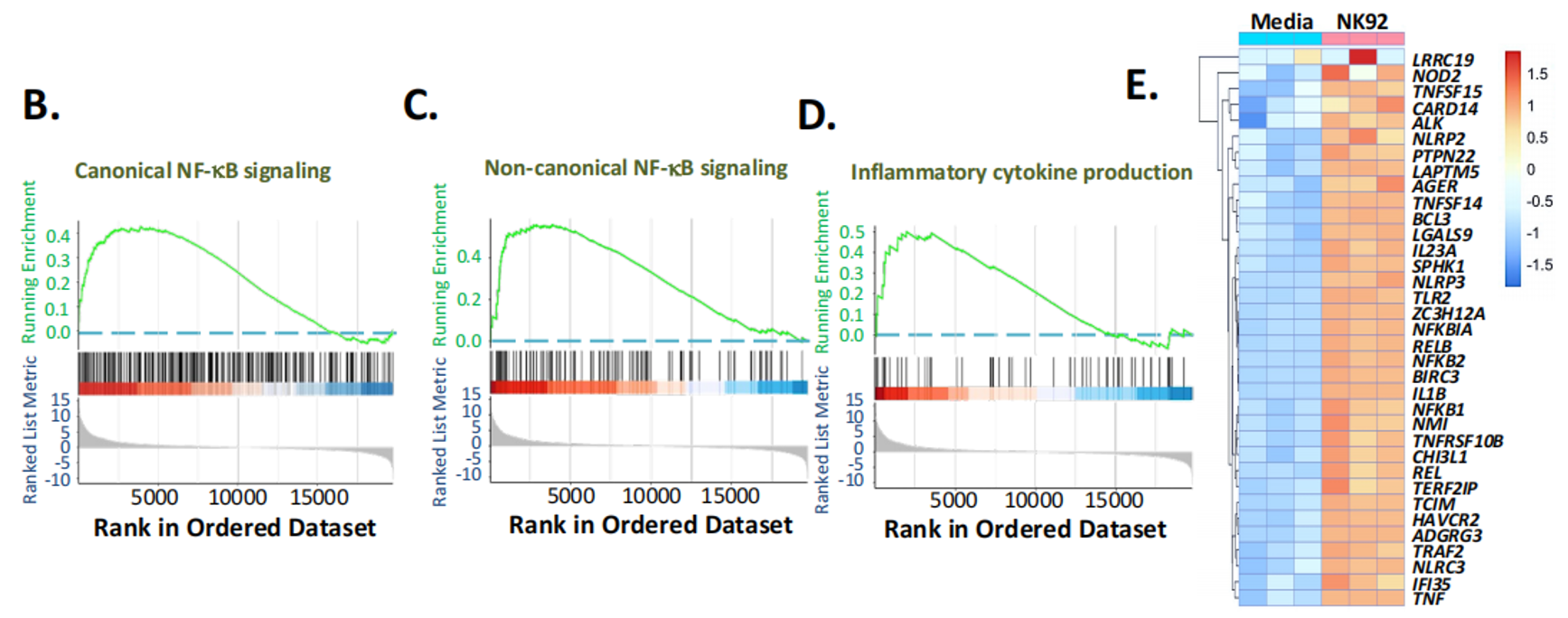
(B-D) Enrichment of NF-κB and inflammatory response pathways, and in the NK high-activated group of TCGA (F) and CGGA (G) GBM cohorts.
Module 3: TFPI2 suppresses GBM proliferation through the POU2F2–CCND1 axis
Approach: Stable TFPI2 knockdown and rescue cells; CCK8, EdU, 3D spheroid, and flow cytometric cell cycle assays; transcriptomic screening of cell cycle genes; TFPI2+POU2F2 double knockdown rescue validation.
Results:
- TFPI2 knockdown increased cell viability, enlarged 3D spheroids, and increased EdU-positive proliferating cells; TFPI2 rescue reversed the phenotype;
- TFPI2 loss upregulated POU2F2 and cyclin CCND1; double knockdown eliminated the abnormal S-phase proliferation;
- Exogenous TFPI2 protein downregulated POU2F2/CCND1 in a dose-dependent manner.
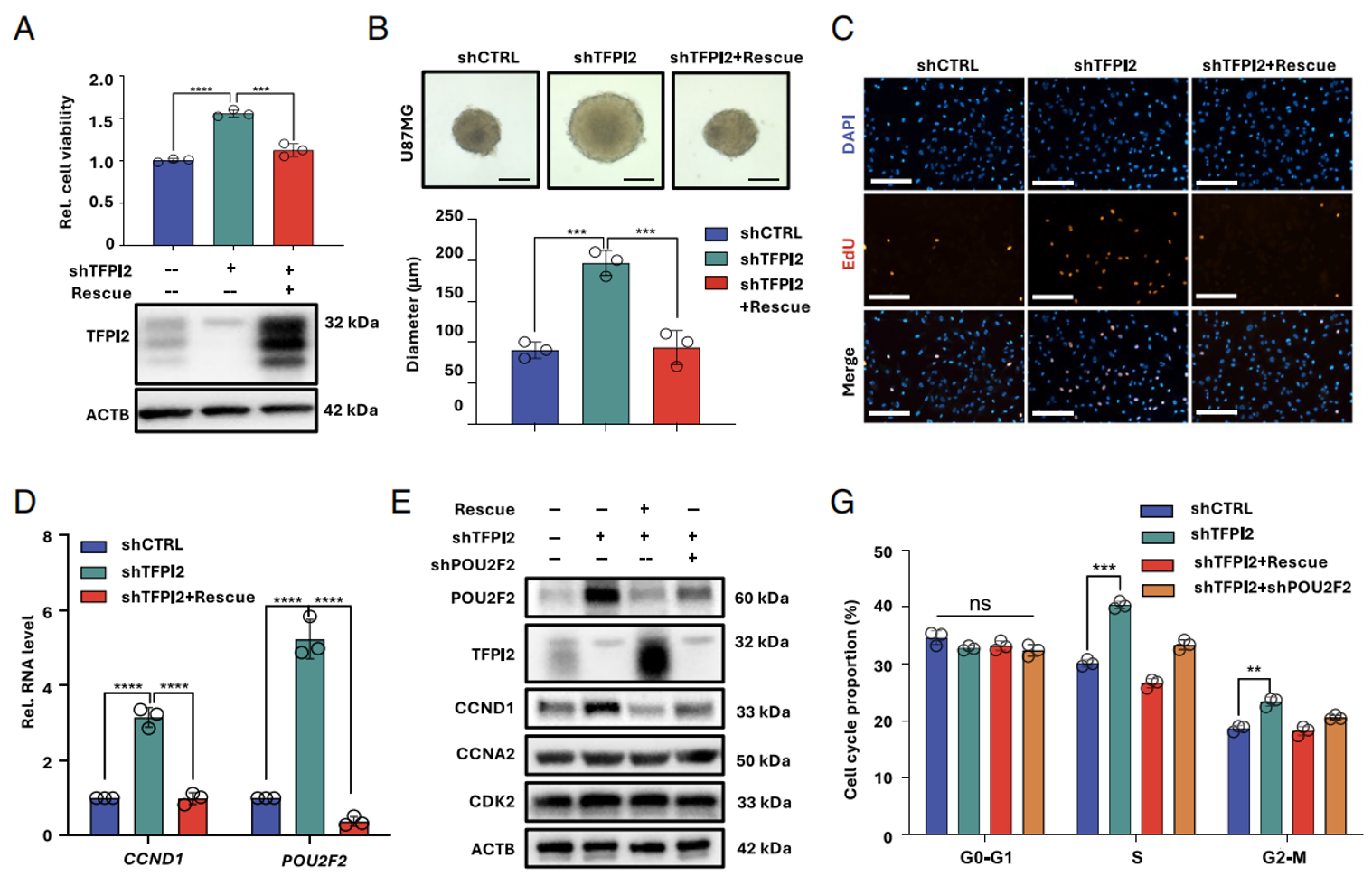
Fig. 3. TFPI2 suppresses GBM proliferation through the POU2F2–CCND1 axis. (A) TFPI2 protein levels and cell viability in TFPI2-knockdown and rescue U87MG cells (Western blotting and CCK-8). (B) Tumor spheroid formation for 7 d (scale bar, 200 μm). (C) EdU incorporation (scale bar, 200 μm). (D) POU2F2 and CCND1 mRNA levels by RT-qPCR. (E) Protein expression of POU2F2, CCND1, CCNA2, and CDK2. (F, G) Cell cycle distribution analyzed by PI staining and FACS.
Module 4: TFPI2 stabilizes LGALS1 to upregulate ICAM1, promoting NK–tumor adhesion and killing
Approach: NK killing and adhesion assays after TFPI2 knockdown; transcriptomic identification of ICAM1; Co-IP mass spectrometry to screen for LGALS1; ubiquitination experiments to clarify the molecular mechanism; LGALS1 knockdown/rescue to validate the axis.
Results:
- TFPI2 knockdown significantly weakened 2D and 3D spheroid NK killing and greatly reduced NK cell adhesion to tumor spheroid surfaces;
- TFPI2 loss downregulated ICAM1; ICAM1 overexpression restored NK sensitivity of TFPI2-knockout cells;
- TFPI2 and LGALS1 co-localized and directly bound intracellularly; TFPI2 loss promoted LGALS1 ubiquitination and degradation, rescued by MG132;
- LGALS1 knockdown directly downregulated ICAM1, reproducing the TFPI2-loss phenotype.
Absin abs9101 usage note: In this module, human primary PB-NK isolation and expansion required abs9101: after PBMC isolation from healthy peripheral blood, abs9101 lysed erythrocytes and removed debris to obtain highly active NK cells for co-culture and fluorescence adhesion imaging — the foundational sample preparation reagent for all in vitro functional experiments in this module.
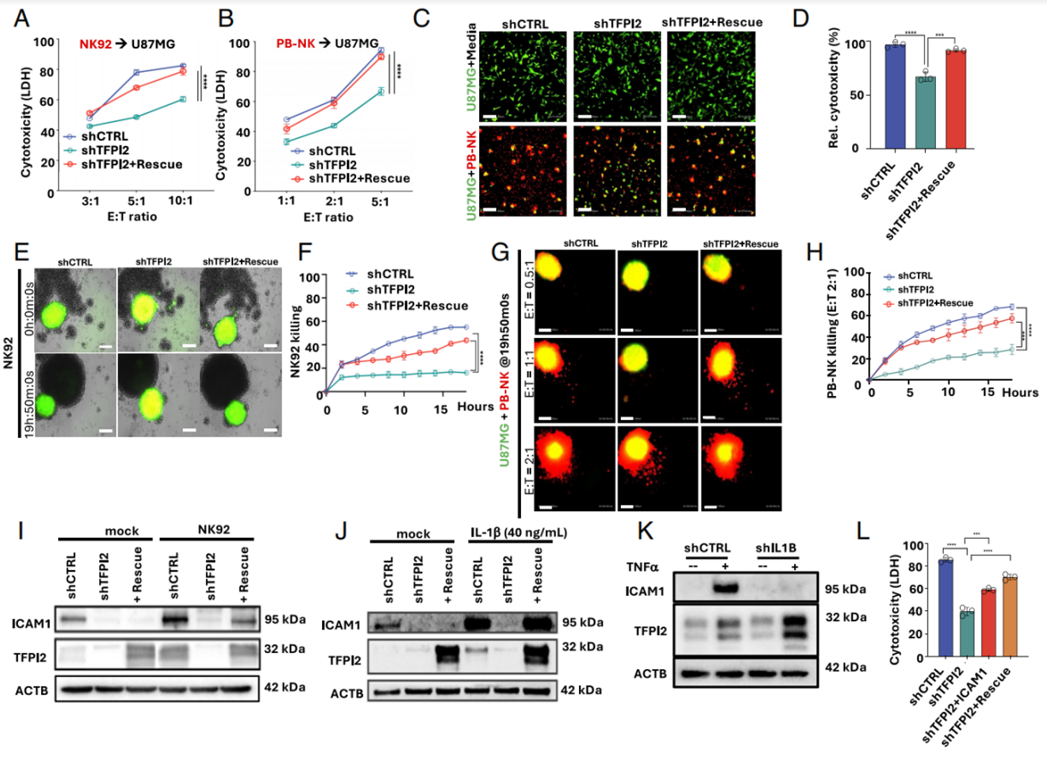
Fig. 4. TFPI2 enhances NK cell–mediated cytotoxicity by supporting ICAM1 expression. (A, B) Cytotoxicity of NK92 cells and PB-NK cells against TFPI2-knockdown U87MG cells by LDH assay. (C, D) PB-NK-mediated killing of EGFP-U87MG cells (scale bar, 200 μm). (E, F) NK92-mediated killing of 3D-cultured EGFP-U87MG spheroids. (G, H) PB-NK-mediated killing of EGFP-U87MG spheroids. (I) ICAM1 protein under TFPI2 modulation with NK92 stimulation. (J) ICAM1 protein with or without IL1β stimulation. (K) ICAM1 and TFPI2 protein in IL1B-knockdown cells treated with recombinant TNFα. (L) ICAM1-encoding lentivector-infected cells challenged by NK92 cells; cytotoxicity measured by LDH assay.
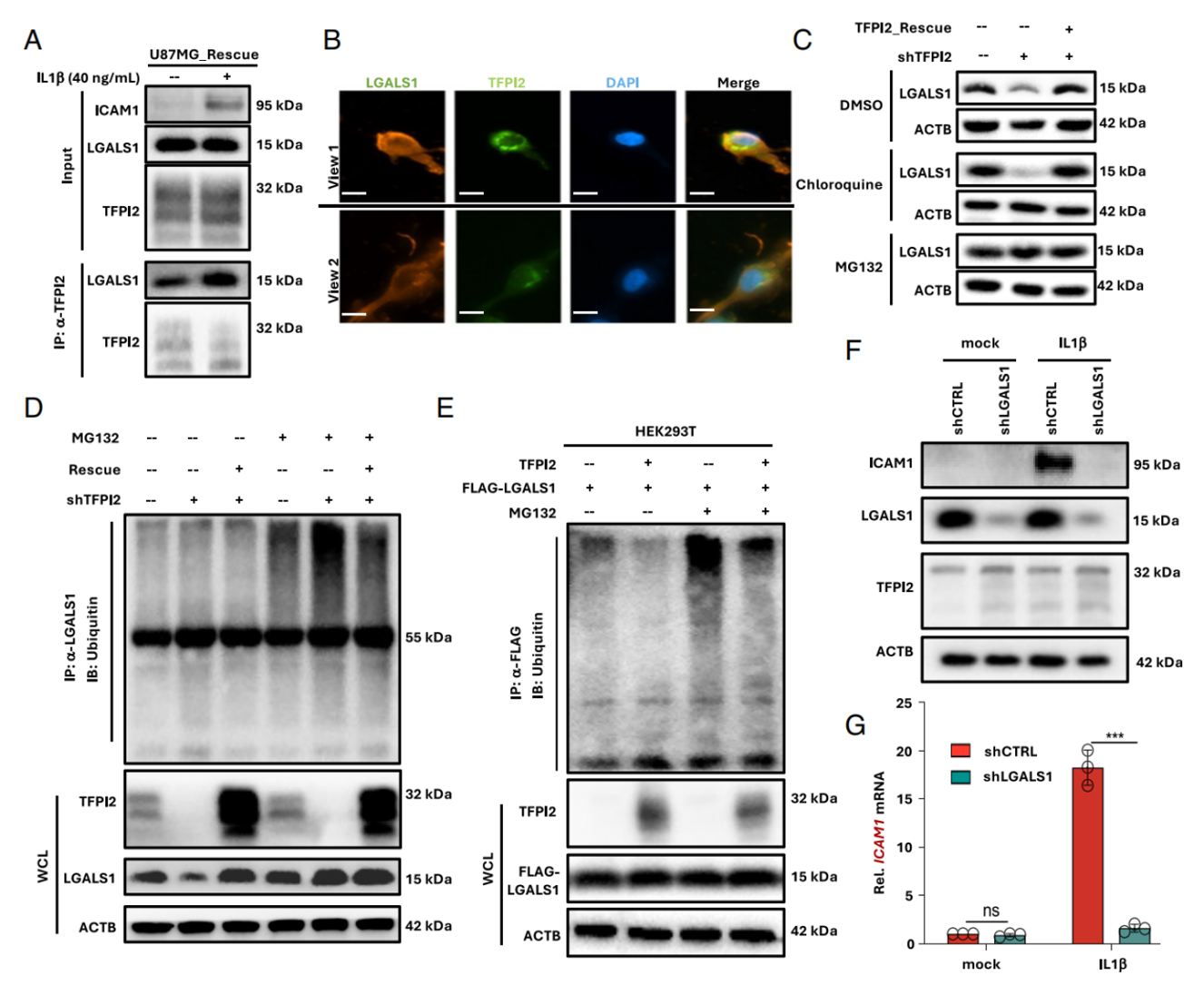
Fig. 5. LGALS1 is required for optimal ICAM1 expression and is stabilized by TFPI2 interaction. (A) Enhanced endogenous TFPI2–LGALS1 interaction upon IL1β stimulation. (B) Colocalization of TFPI2 and LGALS1 (scale bar, 5 μm). (C) LGALS1 protein after MG132 or chloroquine treatment. (D) Polyubiquitination of LGALS1 in TFPI2-knockdown cells. (E) Ubiquitination of LGALS1 in TFPI2-overexpressing HEK293T cells. (F, G) ICAM1 protein and mRNA upon IL1β stimulation and LGALS1 knockdown.
Module 5: TFPI2 selectively suppresses SIGLEC15, relieving NK immunosuppression
Approach: Pan-immune-checkpoint screening after TFPI2 knockdown; recombinant SIGLEC15 treatment to test NK killing and IFNγ secretion; TFPI2+SIGLEC15 double knockout rescue.
Results:
- TFPI2 loss upregulated only SIGLEC15, with no change in PD-L1 expression;
- Recombinant SIGLEC15 treatment significantly reduced NK-mediated tumor lysis and IFNγ secretion;
- Simultaneous knockout of TFPI2 and SIGLEC15 restored NK IFNγ secretion and cytotoxicity.
Absin abs9101 usage note: All in vitro PB-NK cells were purified by abs9101 red cell lysis and expanded for LDH killing and IFNγ ELISA assays.
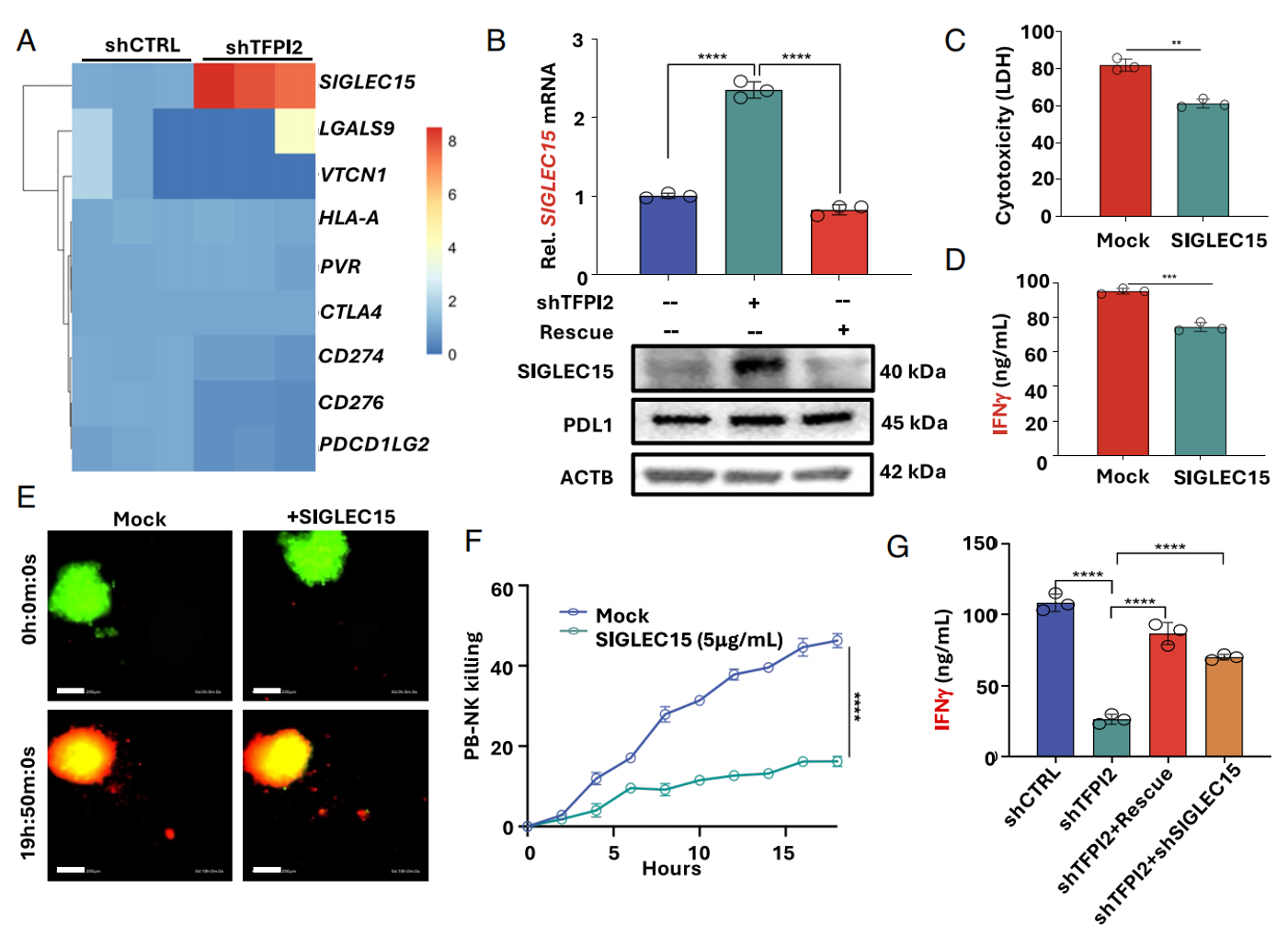
Fig. 6. TFPI2 promotes NK cell activity by selectively suppressing the immune checkpoint SIGLEC15. (A) Heatmap of immune checkpoint molecules in control and TFPI2-knockdown U87MG cells. (B) SIGLEC15 mRNA and protein in knockdown and rescue cells. (C) LDH assay of U87MG cells treated with recombinant SIGLEC15 and NK92 cells. (D) IFNγ secretion measured by ELISA. (E, F) PB-NK-mediated killing of 3D-cultured spheroids treated with recombinant SIGLEC15 (scale bar, 200 μm). (G) IFNγ secretion under single or dual knockdown conditions.
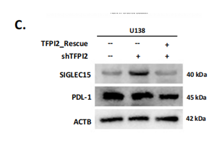
(C) Protein expression of SIGLEC15 and PD-L1 in TFPI2-knockdown and rescue U138MG cells.
Module 6: Multi-level in vivo models validating NK-dependent tumor suppression by TFPI2
Approach: Three intracranial tumor models: immunocompetent GL261, immune-cold CT2A, and NCG humanized U87 xenografts; NK depletion and human PB-NK adoptive transfer groups; mouse brain tissue digestion for TIL extraction and flow cytometric quantification of NK infiltration.
Results:
- GL261 model: Tfpi2 knockdown increased tumor fluorescence burden and shortened survival; no additional tumor promotion after NK depletion; intracranial CD45⁺CD3⁻NK1.1⁺ NK infiltration significantly reduced;
- CT2A cells did not respond to IL1β/TNFα, and Tfpi2 knockdown did not alter tumor growth;
- NCG mice infused with abs9101-purified human PB-NK: tumors clearly shrank in the control group, while the TFPI2 knockout group derived no therapeutic benefit.
Absin abs9101 usage notes (core application scenarios):
- Mouse tumor TIL extraction: intracranial GL261/CT2A brain tissues were enzymatically digested into single-cell suspensions, and abs9101 lysed tissue erythrocytes to remove hemoglobin interference with flow fluorescence, ensuring accurate NK cell gating for quantifying tumor-infiltrating NK proportions;
- Human adoptive NK preparation: after PBMC isolation from healthy peripheral blood, abs9101 removed erythrocytes; purified NK cells were expanded and injected via the tail vein into NCG mice — an essential preprocessing reagent for human immune reconstitution in vivo;
- All mouse spleen and peripheral blood flow samples were preprocessed with a standardized abs9101 red cell lysis workflow, ensuring consistency across multi-group animal experiments and reducing experimental error.
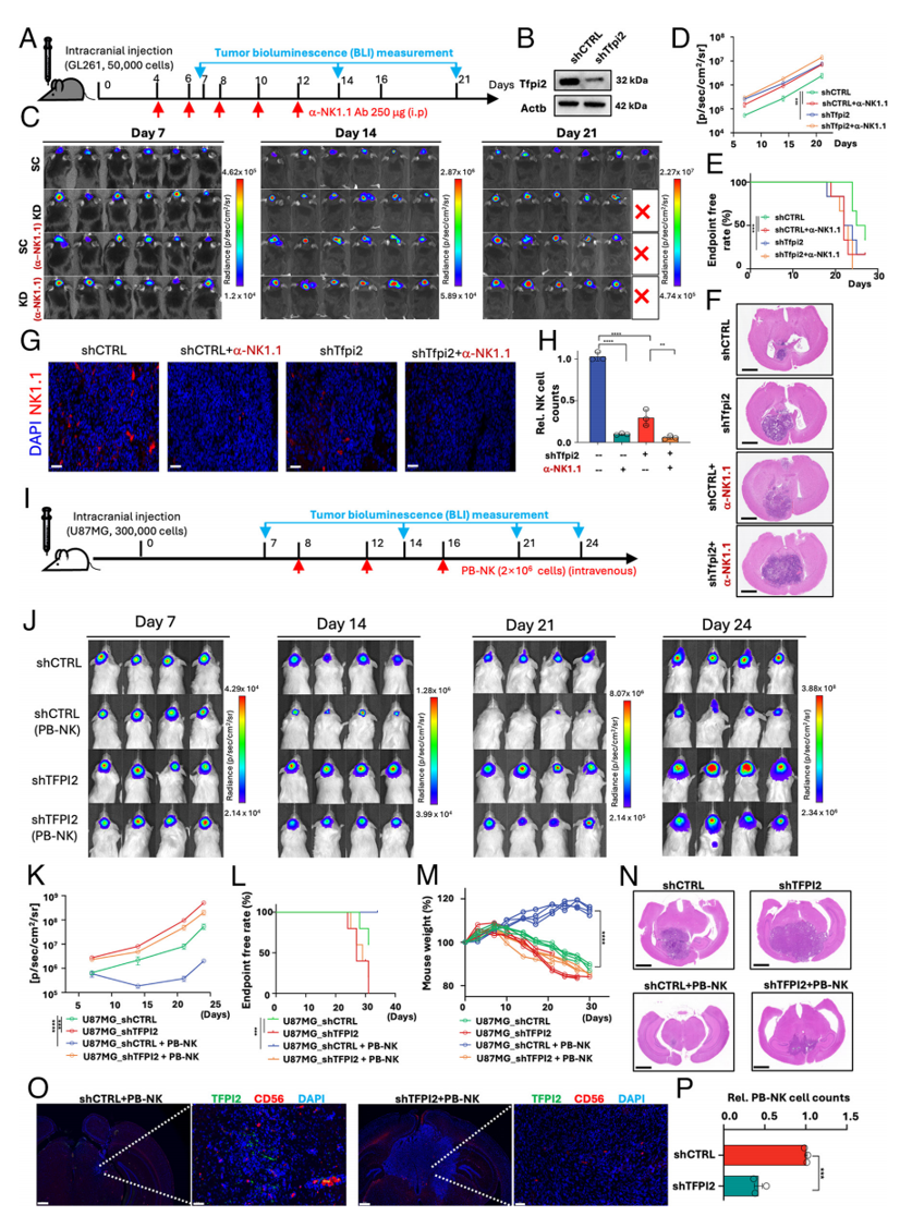
Fig. 7. TFPI2 promotes NK-mediated GBM killing in vivo. (A) Experimental timeline for intracranial implantation and anti-NK1.1-mediated NK depletion in C57BL/6 mice. (B) Tfpi2 knockdown in GL261-Luc cells. (C) Representative bioluminescence images. (D) Quantification of bioluminescence flux. (E) Endpoint-free rate over 27 d. (F) H&E-stained brain sections (scale bar, 2 mm). (G, H) Immunofluorescence staining of NK cells (scale bar, 50 μm). (I) Timeline for human U87MG-Luc implantation and human primary NK transfer in NCG mice. (J) Representative bioluminescence images. (K) Quantification of flux. (L) Endpoint-free rate over 32 d. (M) Body weight changes. (N) H&E-stained brain sections. (O, P) Immunofluorescence staining of human NK cells (CD56) and TFPI2.
V. Overall Summary of Absin abs9101 Red Blood Cell Lysis Buffer in This Study
Absin abs9101 served as the standardized preprocessing core reagent throughout all primary immune cell experiments in vitro and in vivo, providing foundational sample assurance for all NK cell-related functional data. Its roles fall into four aspects:
- Support for human primary NK preparation: lysis of erythrocytes after PBMC isolation from healthy peripheral blood, removing debris and hemoglobin impurities while fully preserving NK cell viability for all in vitro functional experiments including co-culture, 3D spheroid killing, and cytokine secretion assays;
- Standardization of mouse tumor immunophenotyping by flow cytometry: uniform abs9101 red cell lysis of intracranial glioma tissues and mouse peripheral/spleen single-cell suspensions eliminated flow background noise from erythrocytes and accurately quantified the proportion of brain-infiltrating NK cells, supporting GL261/CT2A model heterogeneity comparisons;
- Quality control of adoptive cell therapy in vivo: human NK cells purified with abs9101 maintained stable viability and reliably reconstituted human antitumor immunity after transfer into immunodeficient NC mice, ensuring reproducibility of in vivo efficacy comparisons;
- Experimental stability advantages: the mild abs9101 formulation lyses only anucleate erythrocytes without damaging NK, T, or other target immune cells, accommodating cell culture, flow cytometry, and in vivo transfer scenarios, unifying human/mouse sample preprocessing, reducing experimental variables, and underpinning key conclusions on immune phenotype, killing function, and survival.
Contact Absin
Absin provides antibodies, proteins, ELISA kits, cell culture, detection kits, and other research reagents. If you have any product needs, please contact us.
| Absin Bioscience Inc. worldwide@absin.cn |
 Follow us on Facebook: Absin Bio |