

- Cart 0
- English
The "Immunosuppressive Drivers" in the Tumor Microenvironment: Functions and Therapeutic Potential of Four Key Cytokines
July 08, 2026
Clicks:356


Tumor initiation and progression stem not merely from the aberrant proliferation of cancer cells themselves, but also rely heavily on support from their unique living niche—the tumor microenvironment (TME). Within this complex ecosystem, immunosuppressive cytokines act as key drivers. By regulating immune cell functions and remodeling immune landscapes, they construct a "safe haven" for tumor cells to evade host immune surveillance. Among them, four major cytokines, IL-10, TGF-β, IL-4 and IL-35, serve as core targets for cancer research and therapeutic development owing to their broad immunosuppressive effects and dominant roles across multiple malignancies.

Core Mechanisms of Immunosuppressive Cytokines
Immunosuppressive cytokines in the TME are mainly secreted by immunoregulatory cells (including MDSCs, TAMs, Tregs and Bregs) and tumor cells. Their core function is to disrupt host anti-tumor immune responses via multiple pathways. On one hand, they directly inhibit the proliferation and functional activity of effector immune cells such as cytotoxic CD8+ T cells and natural killer (NK) cells, impairing their tumor-killing capacity. On the other hand, they induce phenotypic and functional transformation of naive immune cells to facilitate the formation of immunosuppressive cell lineages, further amplifying immune suppression. Meanwhile, these cytokines remodel the epigenomic and transcriptional networks of immune cells, alter the growth and proliferative properties of cancer cells, and create favorable conditions for tumor growth, invasion and metastasis.
Properties and Dual Roles of Four Key Cytokines
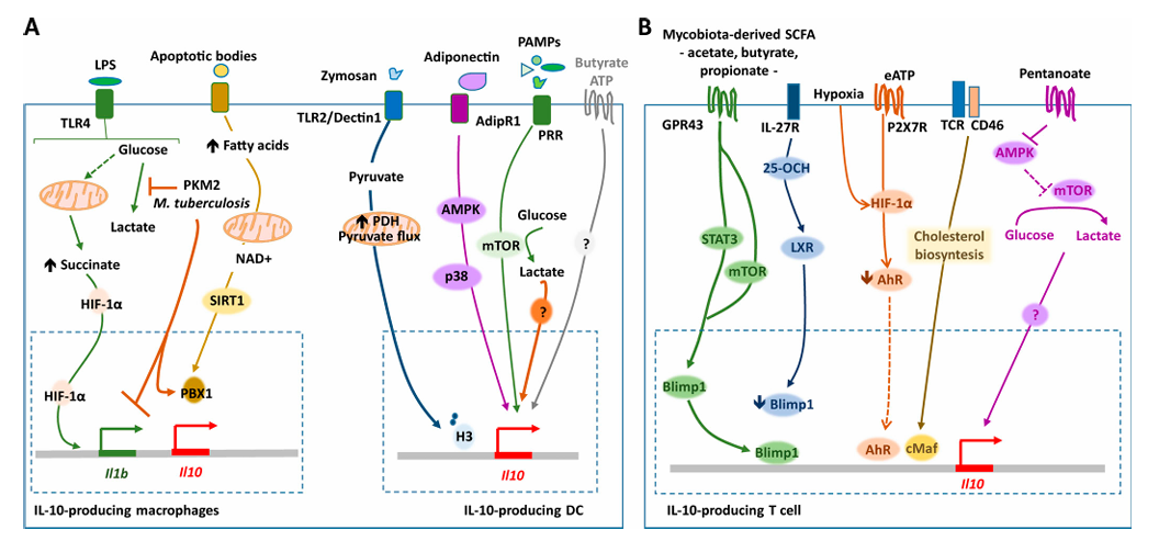
IL-10: A Dual-Function Cytokine with Both Tumor-Promoting and Tumor-Suppressing Potentials
IL-10 is a multifunctional immunomodulatory factor produced by the following cell populations:
- Immune cells: Th2 cells, CD4/CD8 T cells, B cells, macrophages, DCs, neutrophils, mast cells, etc.
- Non-immune cells: Epithelial cells, tumor cells, tissue-resident macrophages (e.g., microglia, cardiac macrophages).
It exhibits distinctive dual functions during tumor progression. In its tumor-promoting capacity, IL-10 downregulates the expression of major histocompatibility complex (MHC) on the surface of cancer cells and antigen-presenting cells (APCs), blocking antigen-specific T cell recognition of tumor cells and thereby suppressing cytotoxic T cell activation. However, under specific conditions, high-dose IL-10 conversely enhances the proliferation and cytotoxic activity of CD8+ T cells while inhibiting pro-tumor inflammatory responses, exerting anti-tumor effects.
This characteristic opens new avenues for therapeutic development. The cetuximab-based IL-10 fusion protein (CmAb-IL10) exerts potent anti-tumor activity by blocking dendritic cell-mediated apoptosis of tumor-infiltrating CD8+ T cells. Its combination with immune checkpoint blockers markedly improves anti-tumor immunity in mice with advanced tumors. These findings suggest that therapeutic strategies targeting IL-10 require flexible inhibition or activation of its pathway based on specific cancer types and patient subgroups.
TGF-β: A Stage-Dependent Functional Switcher in Tumor Progression
TGF-β serves as a core molecule within the immunosuppressive network, broadly inhibiting immune responses through diverse mechanisms: it attenuates the differentiation and function of Th1, Th2 cells and cytotoxic T lymphocytes (CTLs), modulates the abundance and function of regulatory T cells (Tregs) to reinforce immune tolerance, and regulates thymic and peripheral T cell development. Similar to IL-10, TGF-β displays stage-dependent dual effects throughout tumor progression.
At early tumor stages, TGF-β induces apoptosis and suppresses proliferation of multiple cell types including cancer cells, exerting anti-tumor effects. Yet at advanced stages, its function switches: it acts as a critical tumor-promoting factor by regulating genomic instability, epithelial-mesenchymal transition (EMT), angiogenesis, immune evasion and metastasis. Once its pro-tumor functions predominate, TGF-β continuously drives tumor progression. This property dictates that TGF-β-targeted therapies must precisely match the developmental stage of tumors.
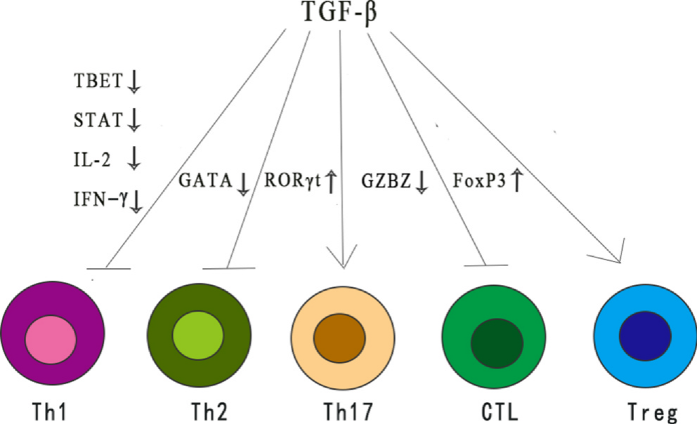
TGF-β regulates distinct T cell subsets via divergent mechanisms and mediates immunosuppression by modulating various immune cell populations.
IL-4: A Master Switch Governing Immune Phenotypes
IL-4 is mainly secreted by Th2 cells via autocrine signaling, with minor production from basophils and mast cells. Its core function is to drive lineage-specific differentiation of Th2 cells and orchestrate humoral immune responses.
- Effects on B cells
Class switch recombination: Induces B cells to switch antibody isotypes from IgM to IgE and IgG1 (in mice; the human counterpart is IgG4). IgE production constitutes a central event in type I hypersensitivity reactions.
Proliferation and survival: Promotes B cell proliferation and viability.
MHC class II expression: Upregulates surface MHC class II molecules on B cells to enhance their antigen presentation capacity.
- Effects on T cells
Promotion of Th2 differentiation: IL-4 is the master cytokine driving naive CD4+ T cell differentiation into Th2 cells. This forms a positive feedback loop: low levels of IL-4 produced by naive T cells drive their own Th2 polarization, while mature Th2 cells massively secrete IL-4, IL-5 and IL-13.
Inhibition of Th1/Th17 differentiation: Suppresses IFN-γ and IL-12 signaling via the STAT6 pathway, thereby blocking Th1 and Th17 cell differentiation.
- Effects on macrophages
Within the TME, IL-4 exacerbates immunosuppression and impairs host tumor immune surveillance by boosting the generation of immunosuppressive M2-type macrophages and myeloid-derived suppressor cells (MDSCs).
Multiple animal studies have validated the therapeutic potential of IL-4 blockade. In mouse models of colon and breast cancer, IL-4 inhibition reduces M2 polarization and MDSC accumulation, restores tumor-specific CD8+ T cell responses, and potentiates anti-OX40 immunotherapy. In non-small cell lung cancer models, IL-4 blockade elevates IL-12 secretion by tumor antigen-specific dendritic cells, augments effector T cell infiltration and proliferation, and effectively alleviates tumor burden. In anaplastic thyroid carcinoma mouse models, IL-4 deletion markedly enhances anti-tumor immunity and overall survival without obvious toxicities. Collectively, these studies identify IL-4 as a promising target for tumor immunotherapy.
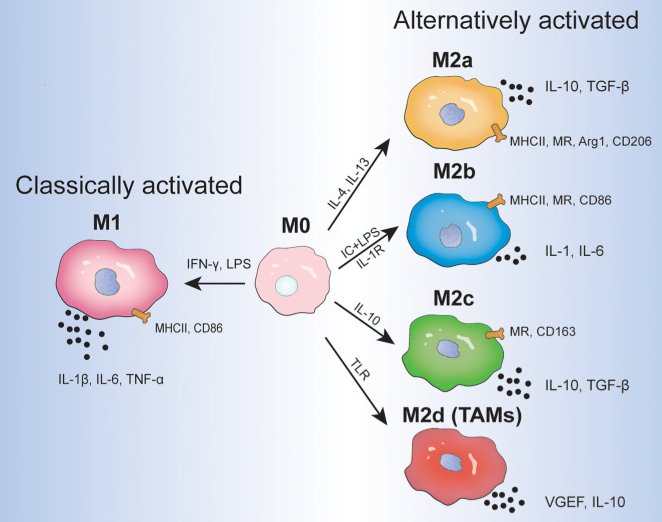 Macrophage phenotypes under different stimuli
- Effects on other cell types
Dendritic cells (DCs): Combined IL-4 and GM-CSF induces monocyte differentiation into dendritic cells.
Epithelial cells: Synergizes with IL-13 to trigger goblet cell metaplasia and excessive mucus secretion in airway epithelia.
Fibroblasts: Stimulates collagen synthesis and participates in tissue fibrosis.
 Induction of monocyte differentiation into dendritic cells
IL-35: A Newly Identified Critical Player in the Immunosuppressive Network
IL-35 is a recently discovered heterodimeric cytokine belonging to the IL-12 family, composed of p35 and EBi3 subunits. Initially confirmed to be predominantly produced by Tregs, its expression in tumor cells has also been verified in recent years. IL-35 plays vital roles in the progression of multiple benign and malignant tumors, including hepatocellular carcinoma, advanced breast cancer, pancreatic ductal adenocarcinoma and non-small cell lung cancer.
Its tumor-promoting mechanisms mainly include: accelerating tumor growth, progression and metastasis by boosting secretion of cytokines such as IL-6 and granulocyte colony-stimulating factor (G-CSF); inhibiting the production of anti-tumor cytokines like IFN-γ to establish an immunosuppressive microenvironment; mediating crosstalk between tumor cells and surrounding immune cells to restrict infiltration and proliferation of tumor-infiltrating lymphocytes (TILs). The discovery of multiple IL-35+ immune cells including M1-type tumor-associated macrophages (TAMs) and DCs demonstrates that tumor-derived IL-35 exerts pro-tumor effects across diverse cellular contexts, making it a vital component of the TME immunosuppressive network.
The cGAS-STING signaling pathway has emerged as a promising target for cancer immunotherapy. Nevertheless, STING agonists exhibit dual-edged sword properties. While STING agonists boost anti-tumor T cell activity, they also elicit pro-tumor effects by driving IL-35-producing regulatory B cells (Bregs), reducing NK cell density and fostering immune suppression. To address these challenges, this study constructed a tumor microenvironment-responsive hollow mesoporous nanosystem that degrades under high glutathione concentrations to release the STING agonist MSA-2 and manganese ions. This nanoplatform facilitates MRI-guided chemodynamic therapy and radiosensitization, potently activating the cGAS-STING pathway via mitochondrial and nuclear DNA damage. Notably, co-administration of this nanosystem with anti-IL-35 blocking agents successfully alleviates Breg-mediated NK cell suppression, restores innate immune responses, and amplifies anti-tumor therapeutic efficacy. This study highlights the pivotal function of anti-IL-35 strategies in reversing immune suppression, reinvigorating innate immunity, and building synergistic theranostic platforms for MRI-guided cancer radioimmunotherapy.
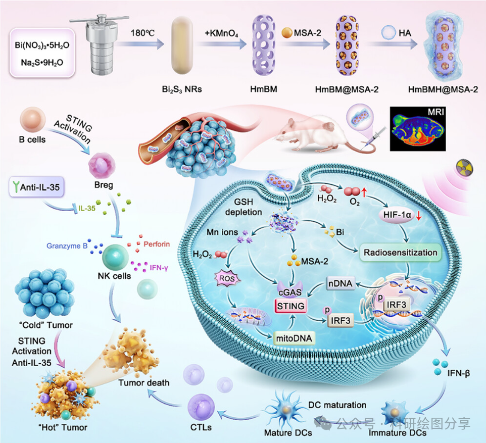
Therapeutic Advances and Prospects Targeting Immunosuppressive Cytokines
Given the central roles of immunosuppressive cytokines in tumor immune escape, cytokine neutralization or depletion represents a vital cancer therapeutic strategy. Such approaches hold potential to overcome poor response to immune checkpoint inhibitors caused by TME-mediated immune suppression, and sensitize tumors to conventional and immunotherapies.
Multiple TGF-β-targeted agents have entered clinical trials. The small-molecule TGF-βRI kinase inhibitor galunsertib (LY2157299) combined with gemcitabine significantly improves overall survival (OS) compared with placebo plus gemcitabine in patients with pancreatic ductal adenocarcinoma (PDAC). The humanized monoclonal antibody GC-1008 (fresolimumab) neutralizes all TGF-β isoforms. The bifunctional molecule M7824 (bintrafusp alfa) simultaneously targets PD-L1 and sequesters TGF-β within the TME. The anti-GARP antibody ABBV-151 markedly attenuates pulmonary metastasis of breast cancer cells by blocking GARP-LAP binding. The genetically modified vaccine belagenpumatucel-L (Lucanix) and antisense oligonucleotide trabedersen (AP12009) downregulate TGF-β2 expression and confer favorable survival benefits in specific patient subgroups.
While the development of targeted therapeutics for IL-10, IL-4 and IL-35 lags behind, encouraging progress has been achieved in basic research and early exploratory studies. Combined regimens of IL-10 fusion proteins with immune checkpoint inhibitors, as well as synergistic combinations of IL-4 blockers with existing immunotherapies, display promising therapeutic value. With deeper mechanistic insights into IL-35, targeted agents against its subunits or receptors will emerge as a key direction for future drug development.
In summary, IL-10, TGF-β, IL-4 and IL-35 are core immunosuppressive cytokines within the tumor microenvironment. Their complex dual functions and pivotal roles in tumor progression provide diverse candidate targets for cancer treatment. In the future, further dissection of the regulatory networks governing these cytokines and optimization of personalized therapeutic regimens will enable cytokine-targeted therapies to be combined with immune checkpoint blockade, chemotherapy, radiotherapy and other modalities, delivering novel therapeutic breakthroughs for cancer patients and ultimately enabling effective control of tumor growth and metastasis.
References:
[1] Roles of TGF-β signaling pathway in tumor microenvironment and cancer therapy. Int Immunopharmacol. 2020 Oct 21;89(Pt B):107101.
[2] Nanosensitizer for Cancer Radioimmunotherapy via Anti‐IL‐35 Blockade Boosted Innate Immunity Activation. Adv. Sci. 2025 June 26;DOI : 10.1002/advs.202504252
Contact Absin
Absin provides antibodies, proteins, ELISA kits, cell culture, detection kits, and other research reagents. If you have any product needs, please contact us.
| Absin Bioscience Inc. worldwide@absin.cn |
 Follow us on Facebook: Absin Bio Follow us on Facebook: Absin Bio |