- Cart 0
- English
Uncovering the Key Axis of PTMC Progression via Single-Cell Combined with Spatial Transcriptomics, Absin Multiplex Fluorescence Kit Empowers Protein Localization Research
July 03, 2026
Clicks:84
Article Title: PROS1‐MERTK Axis Drives Tumor Microenvironment Crosstalk and Progression in Papillary Thyroid Microcarcinoma
Journal: Advanced Science (IF 14.1)
DOI:https://doi.org/10.1002/advs.202413474
Core Reagent:6-color Multiplex Immunofluorescence IHC Staining Kit (Mouse & Rabbit Universal Secondary Antibody) (abs50014)
I. Research Background: Clinical Bottlenecks of PTMC Require Mechanism Breakthroughs
The incidence of papillary thyroid carcinoma (PTC) keeps rising year by year, among which papillary thyroid microcarcinoma (PTMC, diameter ≤ 1cm) accounts for more than 50%. Most PTMC lesions grow slowly with favorable prognosis (10-year survival rate: 93.5%-97%), yet 0.4%-28.8% of cases suffer rapid progression such as tumor enlargement, lymph node metastasis and extrathyroidal invasion. Clinically, specific biomarkers for identifying progression risks and targeted intervention targets are still lacking.
Moreover, after the concept of "overdiagnosis and overtreatment of PTMC" was put forward in 2013, active surveillance (AS) has replaced surgical resection in Japan, the United States and other countries. Nevertheless, distinguishing indolent PTMC from progressive PTMC remains a tough clinical problem, which also constitutes the core goal of this study: to explore vital molecular mechanisms and potential biomarkers for PTMC progression.
II. Core Research Ideas: Systematic Analysis from "Cell Ecosystem" to "Pathway Mechanism"
The research team adopted the logic of "multi-dimensional sequencing + multi-level verification" to clarify PTMC progression mechanisms step by step, with detailed ideas as follows:
1. Sample Design: Full Coverage of Disease Stages to Guarantee Data Representativeness
A total of 19 surgical specimens obtained from 15 patients were enrolled, covering 4 key groups:
- Normal thyroid tissues (4 cases)
- Non-progressive PTMC (4 cases, no disease progression after 5 years of active surveillance)
- Progressive PTMC (5 cases, accompanied by tumor enlargement / metastasis)
- Advanced progressive PTC (6 late-stage cases)
2. Technical Route: Three Steps of "Sequencing Analysis → Spatial Localization → Functional Verification"
- Single-cell level: scRNA-seq was performed on 146,529 cells to classify 32 cell clusters, illustrating dynamic changes of 6 major cell types including thyroid cancer cells, fibroblasts and immune cells;
- Spatial level: combined with 10x Visium spatial transcriptomics, the distribution of different cell populations in tissues was located to analyze tumor heterogeneity in progressive lesions;
- Cell communication level: CellChat was used to analyze ligand-receptor crosstalk between cells, confirming the PROS1-MERTK axis as the key pathway;
- Protein verification level: Absin multiplex IHC/IF kit (abs50014) was applied to detect target protein expression and co-localization, verifying the cellular origin of the pathway;
- Functional verification level: cell migration/invasion assays and mouse subcutaneous xenograft tumor models validated the pro-progressive effect of the PROS1-MERTK axis;
- In-depth mechanism exploration: SCENIC analysis and dual-luciferase reporter assays identified transcription factors (NFYB/FOXP2) regulating PROS1 as well as downstream pathways (WNT/TGF-β).
III. Key Research Achievements: PROS1-MERTK Axis Acts as the "Accelerator" for PTMC Progression
Through systematic analysis, the team concluded four core findings fully supported by solid data:
1. PTC Progression Is Accompanied by Tumor Microenvironment "Immunosuppression + Fibroblast Enrichment"
scRNA-seq Results: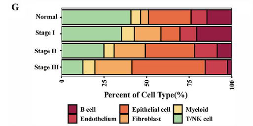
- Along with disease progression (Normal → Stage I → Stage II → Stage III), the proportion of T/NK cells gradually declines with enhanced immunosuppression;
- The percentage of fibroblasts rises remarkably, increasing from approximately 5% in normal tissues to 18% in Stage III lesions, which makes fibroblasts the core "signal sender" in the microenvironment.
2. The PROS1-MERTK Axis Functions as the Critical Hub for Intercellular Communication
CellChat analysis of ligand-receptor interactions between cells revealed that:
- PROS1 (ligand) is mainly secreted by adipogenic cancer-associated fibroblasts (adi-CAFs) in progressive lesions;
- MERTK (receptor) is predominantly expressed on progressive tumor cells and myeloid cells such as tumor-associated macrophages;
- The interaction intensity between the two molecules elevates significantly alongside disease progression (Normal < Stage I < Stage II ≤ Stage III), facilitating tumor progression via paracrine secretion (CAFs → tumor cells) and autocrine secretion (autologous PROS1 secretion by tumor cells).
3. NFYB/FOXP2 Regulates PROS1 Transcription as the "Upstream Switch" of the Pathway
Based on SCENIC transcription factor analysis and dual-luciferase reporter assays: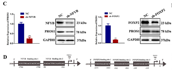
- In fibroblasts, NFYB and FOXP2 can directly bind to the PROS1 promoter to activate its transcription;
- After NFYB/FOXP2 knockdown, the mRNA and protein expression of PROS1 decreases by more than 60%, and the migration capacity of tumor cells is markedly weakened.
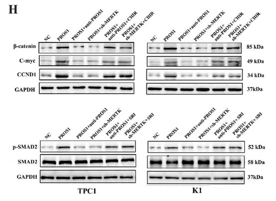
4. The PROS1-MERTK Axis Promotes Progression Through WNT/TGF-β Pathways, and MERTK Can Serve as a Biomarker
Western blot and GSEA analysis confirmed the following results:
 |
 |
- Upon binding to MERTK, PROS1 activates downstream WNT/β-catenin and TGF-β/SMAD2 pathways to boost the proliferation and invasion of tumor cells;
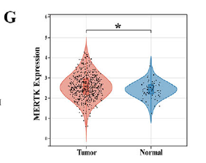
- TCGA database verification demonstrates that MERTK is highly expressed in progressive PTMC/PTC lesions, and patients with high MERTK expression exhibit poorer prognosis with a 35% reduction in disease-free survival, indicating MERTK can act as a potential biomarker and therapeutic target for progressive PTMC.
IV. Empowerment by Absin Products: Multiplex Fluorescence Kit Solves the "Protein Localization Dilemma"
Throughout the whole research, identifying the cellular source of PROS1 and the tissue localization of the PROS1-MERTK axis were core scientific questions. The Absin Multiplex IHC / Immunofluorescence Kit (Cat. No. abs50014) acted as the core tool to address these problems.
1. Product Information & Application Scenarios
| Product Name | Catalog No. | Core Advantages | Application in This Study |
|---|---|---|---|
| 6-color Multiplex Immunofluorescence IHC Staining Kit (Mouse & Rabbit Universal Secondary Antibody) | abs50014 | 1. Compatible with multiple targets (simultaneous detection of 3-4 proteins); 2. High signal specificity with low background; 3. Compatible with laser confocal microscope imaging |
Detection of expression and co-localization of proteins including PROS1, ACTA2 and MERTK in tissue sections |
2. Specific Function in This Research: Direct Confirmation That adi-CAFs Are the Main Source of PROS1
The research team performed multiplex immunofluorescence staining on tissue sections of different disease stages with the abs50014 kit and obtained pivotal evidence: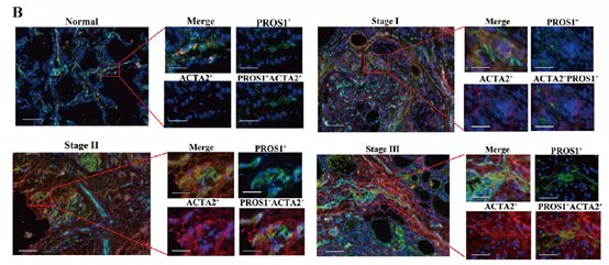
- Staining targets: ACTA2 (fibroblast marker, red), PROS1 (green), DAPI (nucleus, blue);
- Results: In normal tissues and non-progressive PTMC lesions, almost no co-localization was observed between ACTA2+ fibroblasts and PROS1. By contrast, over 80% of ACTA2+ fibroblasts expressed PROS1 in progressive Stage II PTMC and Stage III PTC tissues, with an overlap rate of red-green signals exceeding 75%, which directly verified adi-CAFs as the primary secretory cells of PROS1.
Moreover, this kit was adopted to verify the differential tissue expression of PROS1 and MERTK: the fluorescence signal intensity of PROS1 and MERTK in tumor regions of progressive lesions was 3-5 times higher than that in normal tissues, further supporting the correlation between axis activation and disease progression.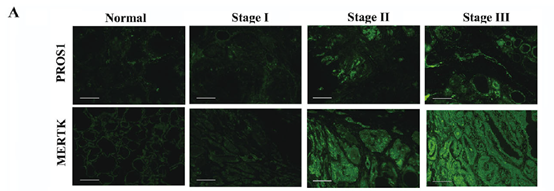

V. Conclusion & Prospect: A Bridge Connecting Basic Research and Clinical Translation
For the first time, this study reveals the central role of the PROS1-MERTK axis in PTMC progression, providing MERTK as a novel potential biomarker and therapeutic target for clinical practice. It also demonstrates the powerful value of the research paradigm combining single-cell sequencing, spatial transcriptomics and multiplex fluorescence validation in the field of cancer microenvironment research.
As a critical verification tool, the Absin Multiplex IHC/IF Kit (abs50014) delivers reliable support for cross-scale mechanism analysis covering protein, cellular and tissue levels, benefiting from its high specificity and multi-channel compatibility advantages. In the future, Absin will keep launching premium research reagents to facilitate more breakthroughs in cancer microenvironment research and precision medicine, laying a solid technical foundation for clinical transformation.
(Note: Original Figure 5B screenshots and detailed product application protocols can be found in the full-text article Advanced Science 2025, Volume 12, e13474, or contact Absin technical support to obtain complete application solutions.)
V. Recommended Absin Multiplex Immunofluorescence IHC Kits
| Catalog No. | Product Name | Specification |
| abs50086 | 2-color Multiplex Immunofluorescence IHC Staining Kit (Anti-Rabbit Secondary Antibody) | 100T |
| abs50087 | 2-color Multiplex Immunofluorescence IHC Staining Kit (Mouse & Rabbit Universal Secondary Antibody) | 100T |
| abs50088 | 3-color Multiplex Immunofluorescence IHC Staining Kit (Anti-Rabbit Secondary Antibody) | 100T |
| abs50089 | 3-color Multiplex Immunofluorescence IHC Staining Kit (Mouse & Rabbit Universal Secondary Antibody) | 100T |
| abs50103 | 3-color Multiplex Immunofluorescence IHC Staining Kit B (Anti-Rabbit Secondary Antibody) | 100T |
| abs50104 | 3-color Multiplex Immunofluorescence IHC Staining Kit B (Mouse & Rabbit Universal Secondary Antibody) | 100T |
| abs50012 | 4-color Multiplex Immunofluorescence IHC Staining Kit (Mouse & Rabbit Universal Secondary Antibody) | 20T/50T/100T |
| abs50028 | 4-color Multiplex Immunofluorescence IHC Staining Kit (Anti-Rabbit Secondary Antibody) | 20T/50T/100T |
| abs50167 | 4-color Multiplex Immunofluorescence IHC Staining Kit B (Mouse & Rabbit Universal Secondary Antibody) | 20T/50T/100T |
| abs50168 | 4-color Multiplex Immunofluorescence IHC Staining Kit B (Anti-Rabbit Secondary Antibody) | 20T/50T/100T |
| abs50013 | 5-color Multiplex Immunofluorescence IHC Staining Kit (Mouse & Rabbit Universal Secondary Antibody) | 20T/50T/100T |
| abs50029 | 5-color Multiplex Immunofluorescence IHC Staining Kit (Anti-Rabbit Secondary Antibody) | 20T/50T/100T |
| abs50014 | 6-color Multiplex Immunofluorescence IHC Staining Kit (Mouse & Rabbit Universal Secondary Antibody) | 20T/50T/100T |
| abs50030 | 6-color Multiplex Immunofluorescence IHC Staining Kit (Anti-Rabbit Secondary Antibody) | 20T/50T/100T |
| abs50048 | 6-color Multiplex Immunofluorescence IHC Staining Kit (Plus) (Anti-Rabbit Secondary Antibody) | 20T/50T/100T |
| abs50049 | 6-color Multiplex Immunofluorescence IHC Staining Kit (Plus) (Mouse & Rabbit Universal Secondary Antibody) | 20T/50T/100T |
| abs50015 | 7-color Multiplex Immunofluorescence IHC Staining Kit (Mouse & Rabbit Universal Secondary Antibody) | 20T/50T/100T |
| abs50031 | 7-color Multiplex Immunofluorescence IHC Staining Kit (Anti-Rabbit Secondary Antibody) | 20T/50T/100T |
| abs50037 | 7-color Multiplex Immunofluorescence IHC Staining Kit (Plus) (Mouse & Rabbit Universal Secondary Antibody) | 20T/50T/100T |
| abs50038 | 7-color Multiplex Immunofluorescence IHC Staining Kit (Plus) (Anti-Rabbit Secondary Antibody) | 20T/50T/100T |
| abs50165 | 7-color Multiplex Immunofluorescence IHC Staining Kit (770 Dye Enhanced Version) (Anti-Rabbit Secondary Antibody) | 20T/50T/100T |
| abs50166 | 7-color Multiplex Immunofluorescence IHC Staining Kit (770 Dye Enhanced Version) (Mouse & Rabbit Universal Secondary Antibody) | 20T/50T/100T |
| abs50018 | 10-color Multiplex Immunofluorescence IHC Staining Kit | 100T |
| abs50083 | Lung Cancer Tumor Microenvironment Multiplex Immunofluorescence IHC Detection Kit (I) | 20T |
| abs50084 | Lung Cancer Tumor Microenvironment Multiplex Immunofluorescence IHC Detection Kit (II) | 20T |
Currently, Absin multiplex staining service is available at a 50% discount, with the price as low as 200 USD per 4-color slide!Click to view details
Acquire Multiplex Protocols
Reply the keyword "Absin Multiplex Protocol" in the official WeChat account "Absin Bioscience" to obtain the complete experimental protocol for multiplex staining.
Contact Absin
Absin provides antibodies, proteins, ELISA kits, cell culture, detection kits, and other research reagents. If you have any product needs, please contact us.
| Absin Bioscience Inc. worldwide@absin.cn |
 Follow us on Facebook: Absin Bio Follow us on Facebook: Absin Bio |