- Cart 0
- English
Unlocking the Secrets of Mitochondrial Research: This mtDNA Extraction Kit Doubles Experimental Efficiency!
May 13, 2026
Clicks:70
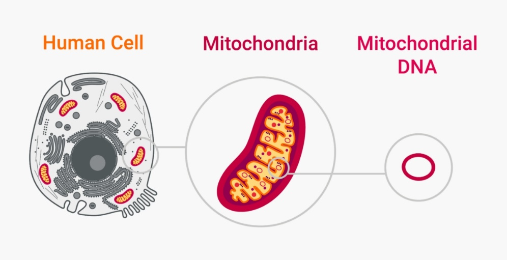
As the intracellular "powerhouse", mitochondria are far more than just energy suppliers. Multiple landmark studies in 2025 have confirmed that mitochondrial dysfunction is closely associated with tumor metastasis, diabetes progression, accelerated aging, and hereditary neuromuscular diseases — cancer cells can enhance metastatic capacity by acquiring mitochondria from neurons, mitochondrial damage repair can reverse pancreatic β‑cell dysfunction, and mutation profiles of mitochondrial DNA (mtDNA) have emerged as novel biomarkers for disease early warning. From molecular evolutionary analysis in basic research to gene therapy exploration for clinical translation, mtDNA is an indispensable core research object.
As circular genetic material independent of the nuclear genome, mtDNA possesses unique features including maternal inheritance, high mutation rate, and high copy number, granting it remarkable research value.
To precisely analyze mtDNA function and variation, the primary prerequisite is high‑purity, nuclear‑DNA‑free, and intact mtDNA samples. Therefore, selecting an efficient and reliable mtDNA extraction protocol is the critical first step for successful mitochondrial research.
Commonly used mtDNA extraction methods currently have respective limitations, failing to balance efficiency, purity and convenience:
‑ Cesium chloride density‑gradient ultracentrifugation: Although high‑purity mtDNA can be obtained, it requires expensive specialized equipment, long centrifugation time, and complicated operation. Moreover, ethidium bromide is carcinogenic. This method is only suitable for fine extraction of small‑scale samples and cannot meet routine experimental requirements.
‑ Triton‑based method: Cells are lysed with non‑ionic detergents, yet nuclear DNA removal is incomplete, causing contamination that impairs accuracy of downstream experiments such as PCR and gene editing, while demanding strict operational skills.
‑ Traditional precipitation method: Cumbersome procedures require repeated phenol‑chloroform extraction using toxic reagents. It is time‑consuming (over 3 hours in total), causes massive mtDNA loss with low yield, and introduces organic reagent contamination.
Addressing these bottlenecks, Absin Mitochondrial DNA Isolation Kit (abs50137) is in stock, applicable to animal tissues and cultured cells. Integrating differential centrifugation and column chromatography core technologies, it balances convenience, high purity and high yield, effectively resolving numerous drawbacks of traditional methods and supporting mitochondrial research.

2. Rapid and efficient, time‑saving: Simplified workflow completes extraction in just over one hour. All supporting reagents are provided in the kit, enabling easy operation even for beginners and greatly improving experimental efficiency.
Step‑by‑step Guide for mtDNA Extraction
Preparation: Add 600 μL (50 tests) or 1100 μL (100 tests) DNase reaction buffer to DNase I, aliquot appropriately and store at ‑20 °C. Mitochondrial lysis buffer is stored at room temperature; dissolve precipitates in a 37 °C water bath if needed. Cool the centrifuge to 4 °C (2‑8 °C). If a refrigerated centrifuge is unavailable, room‑temperature centrifugation is acceptable with 10‑min centrifugation shortened to 5 min, though DNA quality and yield may be affected.
1. Sample Processing
(1) Tissue homogenization: Weigh 100‑200 mg fresh tissue (e.g., liver, brain, cardiac muscle), rinse with PBS or normal saline to remove blood, blot dry with filter paper, mince and place into a small glass homogenizer. Add 1.0 mL ice‑cold cell lysis buffer, and homogenize 20 times on ice at 0 °C;
(2) Cultured cell homogenization: Digest cells, wash with PBS, collect by centrifugation at 800×g for 5‑10 min and count. Use 5×10⁷ cells per extraction. Resuspend cells in 1.0 mL ice‑cold cell lysis buffer, transfer to a small glass homogenizer, and homogenize 30‑40 times on ice at 0 °C;
PS: Perform all steps rapidly at low temperature (manual homogenization) to avoid mitochondrial membrane damage and mtDNA degradation;
2. Transfer tissue or cell homogenate to a centrifuge tube and centrifuge at 1000×g for 5 min at 4 °C;
3. Transfer the supernatant to a new centrifuge tube and centrifuge again at 1000×g for 5 min at 4 °C;
4. Transfer the supernatant to a new centrifuge tube and centrifuge at 12,000×g for 10 min at 4 °C. The post‑centrifugation supernatant contains cytoplasmic components for cytoplasmic protein extraction; transfer supernatant to a new tube, while mitochondria pellet at the bottom;
5. Resuspend mitochondrial pellet in 5 mL mitochondrial wash buffer and centrifuge at 1000×g for 5 min at 4 °C;
6. Transfer the supernatant to a new centrifuge tube and centrifuge at 12,000×g for 10 min at 4 °C. Discard the supernatant; high‑purity mitochondria pellet at the bottom;
7. Resuspend mitochondrial pellet in 100 μL DNase reaction buffer by gentle pipetting, add 10 μL DNase I solution (see Preparation), mix well, and incubate in a 37 °C water bath for 10 min. This step digests nuclear DNA adsorbed on the mitochondrial surface. Centrifuge at 12,000×g for 5 min at 4 °C. Discard supernatant as much as possible, resuspend mitochondrial pellet in 200 μL TE buffer, and centrifuge at 12,000×g for 5 min to remove residual DNase.
8. Resuspend the resulting pellet in 200 μL TE buffer. Add 10 μL RNase A and 200 μL mitochondrial lysis buffer, mix gently (do not pipette vigorously), incubate for 1‑2 min (no more than 5 min), then add 150 μL protein precipitation solution and mix rapidly. Centrifuge at 12,000×g for 5 min at 4 °C. This step further removes nuclear DNA.
9. Transfer the supernatant to a new centrifuge tube (For restriction digestion analysis: perform one extraction with an equal volume of phenol‑chloroform‑isoamyl alcohol (25:24:1), followed by one chloroform extraction, or two sequential chloroform extractions. Generally, this step removes trace proteins and saccharides yet causes mtDNA loss, and can be omitted for PCR without affecting downstream experiments). Add 6× volume of isopropanol (or 2.5× volume of ethanol if isopropanol is unavailable; split into two tubes if overfilled) and 5‑10 μL nucleic acid coprecipitant, mix well, precipitate at ‑20 °C for approximately 30 min (optional), and centrifuge at 12,000×g for 10 min at 4 °C.
10. Discard supernatant, wash with 1 mL 70% ethanol, and centrifuge at 12,000×g for 10 min at 4 °C. Repeat 70% ethanol washing once more.
11. Discard supernatant, centrifuge for an additional 1 min and remove residual supernatant without touching the pellet, air‑dry for 5‑10 min with the lid open.
12. Add 20‑30 μL TE buffer (TE buffer is recommended for long‑term mtDNA storage (> 1 week); ultrapure water can be used for short‑term storage), tap the tube bottom gently, incubate in a 37 °C water bath for 5 min to dissolve mtDNA.
13. Perform DNA electrophoresis analysis, store at ‑20 °C, and proceed to downstream experiments.
PS: mtDNA is inherently low‑abundance. If undetectable by electrophoresis, increase loading volume, dissolve the pellet in a smaller volume of TE buffer, or directly proceed to PCR detection.
From basic research to clinical translation, high‑quality mtDNA samples are the key to breaking research bottlenecks. Featuring precise decontamination, rapidity, convenience, stability and reliability, this high‑efficiency mtDNA extraction kit serves as a powerful assistant for mitochondrial research. It ensures accurate and efficient extractions and credible data, accelerating breakthrough progress in your research!
Recommended Absin Related Products
|
Cat. No. |
Product Name |
Size |
|
Mitochondrial DNA Isolation Kit |
50T/100T |
|
|
Mitochondrial Membrane Potential Assay Kit (JC‑1) |
100T |
|
|
Tissue Mitochondria Isolation Kit |
50T |
|
|
Cell Mitochondria Isolation Kit |
50T |
Contact Absin
Absin provides antibodies, proteins, ELISA kits, cell culture, detection kits, and other research reagents. If you have any product needs, please contact us.
| Absin Bioscience Inc. worldwide@absin.cn |
 Follow us on Facebook: Absin Bio Follow us on Facebook: Absin Bio |