- Cart 0
- English
Progressive Staining Principle and Histological Applications of Mayer's Hematoxylin Stain
May 12, 2026
Clicks:69
Hematoxylin and Eosin (H&E) staining serves as the "gold standard" in histopathological diagnosis, with the precise selection of nuclear dyes being one of its core technologies. Among numerous hematoxylin formulations, Mayer's Hematoxylin Solution has become an essential tool for immunohistochemical counterstaining, fine-needle aspiration cytology, and routine tissue section staining due to its unique progressive staining characteristics. A thorough understanding of its chemical nature and mechanism of action helps researchers obtain clearer and more diagnostically valuable histological images.
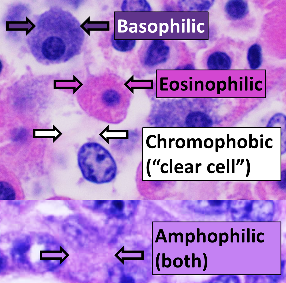
Figure: Staining characteristics of different cellular components in H&E staining. Cell nuclei are blue (basophilic), and cytoplasm is pink to orange-red (acidophilic)
From Logwood to Dye: What is the Chemical Nature of Hematoxylin?
Hematoxylin is not a synthetic dye but a natural compound extracted from the heartwood of the logwood tree (Haematoxylin campechianum). Its staining mechanism relies on oxidation—hematoxylin itself has no staining activity and must be oxidized to haematein in the presence of metal ions (mordants) before it can bind to nucleic acids in tissues.
Aluminum or iron salts are commonly used as mordants, where aluminum mordants produce a blue hue and iron mordants generate a deeper blue-black color. Mayer's formulation is prepared with a low concentration of hematoxylin, which determines its "progressive" staining behavior.
What is the Difference Between Progressive and Regressive Staining?
This is a critical technical classification when selecting hematoxylin solutions. Based on dye concentration and staining intensity, hematoxylin solutions are divided into two systems:
Progressive Staining (Mayer's Type):
With a relatively low dye concentration, it exhibits selective staining properties. It stains only cell nuclei without coloring the cytoplasm within an appropriate staining time. Even with prolonged staining, non-specific cytoplasmic staining does not occur, so clear nuclear staining can be achieved without a differentiation step.
Regressive Staining (Harris, Weigert's Types):
With a high dye concentration, it intensely stains all tissue components (nuclei and cytoplasm). To achieve proper color contrast, differentiation with acid alcohol is mandatory to "decolorize" excess dye bound to the cytoplasm, leaving only nuclear staining. Improper differentiation often leads to faint nuclear staining or residual background in the cytoplasm.
Mayer's formulation is a typical progressive system, making it irreplaceable in scenarios requiring high cytoplasmic integrity.
Which Experimental Scenarios Are Most Suitable for Mayer's Hematoxylin?
This staining solution is applied across multiple pathological and research fields:
Nuclear Counterstaining in Immunohistochemistry:
After DAB chromogenic development or other enzyme-labeled visualization, mild nuclear counterstaining with Mayer's Hematoxylin (usually 1–5 minutes) provides nuclear localization references without masking specific antigen signals. Its lack of cytoplasmic staining avoids confusion with positive chromogenic signals.
Cytological Smears and Fine-Needle Aspiration Specimens:
For cytological samples such as cell blocks and pleural effusion smears, progressive staining ensures clear nuclear visualization while preserving cytoplasmic morphological integrity, facilitating observation of cytoplasmic features and intercellular relationships.
Alternative for Routine H&E Staining:
Although Harris hematoxylin is widely used in standard H&E protocols, Mayer's formulation combined with hydrochloric alcohol differentiation or pure alcohol differentiation can produce unique contrast effects by eliminating cytoplasmic staining in scenarios requiring more cytoplasmic details or special studies.
Bluing of Tissue Sections:
Tissue sections initially appear purple or reddish-purple after hematoxylin staining due to dye color development under acidic conditions. Brief treatment in alkaline solutions (e.g., dilute ammonia water, Scott's tap water substitute, or lithium carbonate solution) converts the dye to its characteristic blue color—a process known as "bluing," which is indispensable for standard blue nuclear staining.
How to Properly Perform the Staining Procedure?
Standard operating procedures are divided into two protocols based on counterstaining methods:
Eosin Counterstaining Protocol (Standard H&E Procedure):
- After deparaffinization and rehydration, incubate sections in Mayer's Hematoxylin Solution for approximately 15 minutes;
- Rinse with warm tap water for 15 minutes (this step also aids bluing);
- Quick rinse with distilled water;
- If using alcohol-soluble eosin, briefly rinse with 95% ethanol (30 seconds) first;
- Counterstain with Eosin Y for 30–60 seconds;
- Dehydrate and clear twice with anhydrous ethanol and xylene, 2 minutes each;
- Mount with resinous medium.
Special Staining Solution Counterstaining Protocol:
- After completing the target staining procedure, rinse with distilled water;
- Incubate in Mayer's Hematoxylin Solution for 1–5 minutes (adjust based on tissue type);
- Rinse with tap water or dilute alkaline solution until cell nuclei turn blue;
- Rinse with distilled water;
- Select water-soluble or alcohol-soluble mounting medium according to the properties of subsequent staining solutions.
What Are the Key Control Points for Successful Staining?
The following technical details are critical for ideal staining results:
Staining Solution Monitoring:
Mayer's Hematoxylin Solution must be filtered before each use to remove oxidized precipitate particles and avoid metal deposits on tissue sections. Repeatedly used solutions gradually lose staining capacity, manifested as a significant prolongation of staining time—replace with fresh solution promptly in this case.
Effect of Water Quality on "Bluing":
Warm tap water (weakly alkaline) can replace dilute alkaline solutions for bluing, shortening the staining process. Note that tap water in some regions is acidic and unsuitable for direct bluing. A simple test: if nuclei remain purple or reddish-brown after bluing, insufficient bluing is indicated, and alkaline solution treatment is required.
Eosin Staining Balance:
Excessive eosin staining may mask nuclear details. Differentiate by prolonging alcohol washing time or using lower-concentration ethanol (e.g., 90% ethanol instead of pure ethanol) to adjust eosin saturation and ensure clear nuclear-cytoplasmic contrast.
Subtle Effects of pH:
The pH environment during staining directly affects the final outcome. Acidic conditions enhance red tones, while alkaline conditions enhance blue tones. If dilute alkaline solution is used before eosin staining, rinse thoroughly with tap water for 2–3 minutes to avoid residual alkalinity interfering with acidic eosin binding.
Interpretation Criteria for Staining Results
Successful Mayer's Hematoxylin staining should exhibit the following features:
- Cell Nuclei: Clear blue with visible nucleolar structures;
- Cytoplasm: Retains pink to orange-red (depending on counterstain), no non-specific hematoxylin staining;
- Erythrocytes: Bright red color;
- Background: Clean, free of dye precipitates or metal salt crystals.
Necessary Preparations Before Use
For first-time use, it is recommended to perform a pilot experiment on 1–2 samples to determine the optimal staining time (usually adjusted within 1–15 minutes). Staining time should be flexibly adjusted based on actual conditions, as tissue processing degrees and fixative types (formaldehyde fixation duration) vary between batches.
After staining, ensure no excess water is introduced into the hematoxylin solution before mounting to avoid dilution. Meanwhile, replace dehydrating ethanol and clearing xylene (or substitutes) daily to maintain gradient dehydration efficiency and prevent section dullness or mounting bubbles.
With the continuous advancement of histopathological technology, Mayer's Hematoxylin Solution provides reliable technical support for precise diagnosis and research observations with its mild progressive staining characteristics. Mastering its principles and operational essentials is a fundamental skill for every histology technician.
Recommended Absin Mayer's Hematoxylin Solution:
| Cat. No. | Product Name | Size |
|---|---|---|
| abs9215 | Mayer's Hematoxylin Solution | 100mL/500mL |
Contact Absin
Absin provides antibodies, proteins, ELISA kits, cell culture, detection kits, and other research reagents. If you have any product needs, please contact us.
| Absin Bioscience Inc. worldwide@absin.cn |
 Follow us on Facebook: Absin Bio Follow us on Facebook: Absin Bio |