- Cart 0
- English
Mitochondrial Targeting and Multidrug Resistance Detection Applications of Rhodamine Dyes
May 11, 2026
Clicks:72
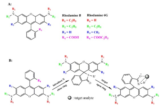
Rhodamine is a family of basic fluorescent dyes with a xanthene core structure, including derivatives such as Rhodamine 123, Rhodamine 6G and Rhodamine B. Featuring unique cationic properties, excellent photostability and tunable spectral characteristics, these dyes are widely applied in cell biology, pharmacology and molecular imaging. From mitochondrial membrane potential detection and P‑glycoprotein efflux‑function evaluation to laser media and quantitative drug analysis, rhodamine dyes have become indispensable tool molecules in modern biomedical research.
Why Do Rhodamines Target Mitochondria?
Mitochondria are the central hubs of cellular energy metabolism, maintaining a negative inner‑membrane potential (Δψm) of approximately −150 to −180 mV. Rhodamine 123 is a cationic dye with positive charge, which actively accumulates in the mitochondrial matrix driven by the electrochemical gradient of the negative inner‑membrane potential. This accumulation property makes it a classic probe for detecting mitochondrial membrane potential (MMP).
When mitochondria are functionally intact with normal membrane potential, Rhodamine 123 concentrates inside mitochondria and emits bright green fluorescence (emission ≈530 nm). During apoptosis or mitochondrial dysfunction, membrane potential collapses (depolarization), releasing the dye and weakening or eliminating fluorescent signals. Based on this principle, Rhodamine 123 is widely used to assess cell viability, monitor early apoptotic events and screen compounds affecting mitochondrial function.
Notably, Rhodamine 123 has relatively low toxicity and does not significantly quench mitochondrial respiration like certain lipophilic cationic dyes (e.g., JC‑1), making it suitable for long‑term live‑cell imaging. However, high concentrations or prolonged incubation may interfere with mitochondrial function; therefore, dye concentration (typically 0.1–10 μg/mL) and incubation time (15–30 min) must be optimized experimentally.
How Does Rhodamine Detect P‑Glycoprotein Function via Flow Cytometry?
P‑glycoprotein (P‑gp/MDR1) and multidrug‑resistance‑associated protein 1 (MRP1) belong to the ATP‑binding‑cassette (ABC) transporter superfamily and play critical roles in tumor multidrug resistance (MDR). These efflux pumps recognize and expel various hydrophobic drugs, reducing intracellular drug concentration and leading to chemotherapy failure.
Rhodamine 123 and Rhodamine 6G are specific substrates of P‑gp and MRP1, serving as convenient probes for efflux‑pump function assays. Typical flow‑cytometry experimental designs include:
Basal efflux assay: Incubate cell suspensions with rhodamine dyes (typically 0.1–1 μM) at 37 °C for cellular uptake. Wash off extracellular dye and incubate further (30–60 min) before detecting intracellular fluorescence intensity. Cells with active P‑gp rapidly efflux the dye, showing low intracellular fluorescence signals.
Inhibition validation assay: After adding P‑gp‑specific inhibitors (e.g., verapamil, cyclosporine A, PSC833), rhodamine efflux is blocked and intracellular fluorescence significantly increases, confirming the specificity of the efflux mechanism.
Applications in Multidrug‑Resistance Mechanism Research
In tumor‑biology research, rhodamine dyes are standard tools for evaluating chemotherapy resistance. Studies show that Rhodamine 123 inhibits clonal growth of cancer cells in vitro, an effect related to its role as a P‑gp substrate — high‑concentration rhodamine may modulate drug sensitivity in resistant cells via competitive binding or transporter interference.
Furthermore, studies linking rhodamine to placental P‑gp reveal barrier mechanisms of drug transport during pregnancy. P‑gp is highly expressed in placental syncytiotrophoblasts, protecting fetuses from potential toxic substances in maternal circulation. Using rhodamine as a model substrate, researchers can assess placental drug permeability and optimize drug safety during gestation.
For kinetic studies of MRP1‑mediated efflux, Rhodamine 6G exhibits unique advantages. Compared with Rhodamine 123, it has higher photostability and better fluorescence quantum yield, making it ideal for quantitative kinetic analysis.
Performance of Rhodamines in Lasers and Imaging
Beyond biological probing, Rhodamine 6G is a representative high‑performance laser dye. Its ethanol solution has a strong absorption peak near 514 nm and a fluorescence emission peak at 550–560 nm (yellow‑green), with excellent photochemical stability and moderate gain bandwidth. It serves as the standard gain medium for argon‑ion‑pumped dye lasers and is widely adopted in laser spectroscopy and photodynamic‑therapy (PDT) light‑source development due to high conversion efficiency and low cost.
In fluorescence microscopy, Rhodamine B is commonly used for multiplex fluorescence labeling owing to its deep‑red fluorescence (emission ≈580 nm) and good photostability. Its spectral profile is well separated from FITC (green) and DAPI (blue), suitable for colocalization studies. Rhodamine B is also applied in cell‑migration tracking (e.g., microsphere labeling), neuronal tracing and fluorescence labeling in materials science.
Applications in Drug Assay and Quality Control
In pharmaceutical analytical chemistry, Rhodamine B is used as a standard reference material for drug quantitation, identification and activity screening. With stable chemical structure and high purity (typically ≥98% HPLC grade), it is suitable as a standard for HPLC or fluorospectrophotometry. In pharmacological experiments, Rhodamine B evaluates the performance of novel fluorescent probes or acts as a positive control to verify detection‑system sensitivity.
Key Technical Notes for Experimental Operation
Solvent selection and solubility: Rhodamine derivatives show significant solubility differences. Rhodamine 123 is soluble in ethanol (20 mg/mL), DMSO and methanol, slightly soluble in water; Rhodamine 6G produces scarlet solutions with green fluorescence in water and red‑yellow fluorescence in alcohols; Rhodamine B dissolves in water and ethanol. Prepare stock solutions in DMSO or ethanol (5–10 mM), aliquot and store at −20 °C protected from light, avoiding repeated freeze‑thaw cycles.
Staining‑condition optimization: For mitochondrial staining, dilute dyes in serum‑free medium or PBS to prevent serum‑protein binding that reduces effective concentration. Thoroughly wash off non‑specifically bound extracellular dye after staining. For live‑cell imaging, use anti‑fade mounting medium.
Detection‑parameter setting: For flow‑cytometry detection of Rhodamine 123/6G, use an excitation wavelength of 488 nm (compatible with the FITC channel) and a detection channel of 530/30 nm (green light). Adjust detector voltage to prevent signal overflow due to high brightness of rhodamine dyes.
Control setup: Unstained cells must be used as negative controls, and P‑gp‑inhibitor‑treated groups as functional validation controls. For mitochondrial‑membrane‑potential detection, use CCCP (carbonyl cyanide m‑chlorophenylhydrazone) treatment as a positive control for complete depolarization.
As precision medicine and drug development increasingly focus on individual resistance profiles, rhodamine dyes serve not only as visualization tools but also as critical probes for analyzing membrane‑transport mechanisms and assessing chemotherapy sensitivity. By rationally selecting derivatives and optimizing experimental conditions, researchers can fully exploit the multidimensional value of these classic dyes in biomedical research.
Recommended Absin Rhodamine Products
| Cat. No. | Product Name | Size |
|---|---|---|
| abs42026664 | Rhodamine 123 | 25 mg / 100 mg |
| abs47050724 | Rhodamine 6G | 100 g |
| abs47002031 | Rhodamine B, AR Grade | 100 g / 500 g |
| abs47006555 | Rhodamine B, HPLC ≥98% | 20 mg |
Contact Absin
Absin provides antibodies, proteins, ELISA kits, cell culture, detection kits, and other research reagents. If you have any product needs, please contact us.
| Absin Bioscience Inc. worldwide@absin.cn |
 Follow us on Facebook: Absin Bio Follow us on Facebook: Absin Bio |