- Cart 0
- English
Why do you always fail to obtain authentic data from your tumor immune cell analysis?
May 11, 2026
Clicks:74
In laboratories engaged in tumor immunology research, a perplexing phenomenon often baffles investigators: identical tumor samples, identical flow cytometry antibody panels, yet drastically varying proportions of immune cell subsets across different experimental batches. Sometimes the proportion of T cells is abnormally low, sometimes the surface antigen expression of myeloid cells is "strangely" diminished, and even single-cell sequencing data reveals a large number of "double-positive" abnormal cells. The root cause often lies not in the analytical techniques themselves, but in the first step of sample preparation—tumor tissue dissociation. Conventional mechanical grinding or single-enzyme digestion either results in cell death and antigen loss, or fails to effectively release infiltrating cells from the tumor microenvironment. Tumor Tissue Dissociation Kits are professional tools designed to address this technical bottleneck.
What exactly is a Tumor Tissue Dissociation Kit?
A Tumor Tissue Dissociation Kit is a tissue digestion system composed of optimally combined biological enzymes, specifically engineered to gently, rapidly, and efficiently enzymolyze solid tumor tissues into single-cell suspensions. The kit is formulated to match the characteristics of tumor tissues from different species (human, rat, mouse). By precisely regulating the specificity and activity of enzymes, it maximally preserves the viability of cell surface antigens and cellular integrity while degrading the extracellular matrix.
Technically, a complete kit system comprises the following core components:
- Tissue Dissociation Enzyme Cocktail: A synergistic formulation of multiple proteolytic enzymes (e.g., collagenase, neutral protease, elastase) targeting the degradation of extracellular matrix components such as collagen, elastin, and fibronectin in the tumor stroma
- Tissue Preservation Solution: Used for the short-term transport and storage of fresh tumor tissues, maintaining tissue viability and reducing cell death caused by ischemia and hypoxia
- Cell Isolation and Purification System: Percoll density gradient centrifugation medium for further purification of dissociated cells and enrichment of specific cell subsets (e.g., tumor-infiltrating lymphocytes)
- Supporting Consumables: 70μm cell strainers (to remove tissue debris), red blood cell lysis buffer (to eliminate red blood cell contamination in tumor tissues), and cell staining buffer (specific for flow cytometry analysis)
This integrated "enzymolysis + purification" design ensures efficient conversion from tumor tissue to high-quality single-cell suspension.
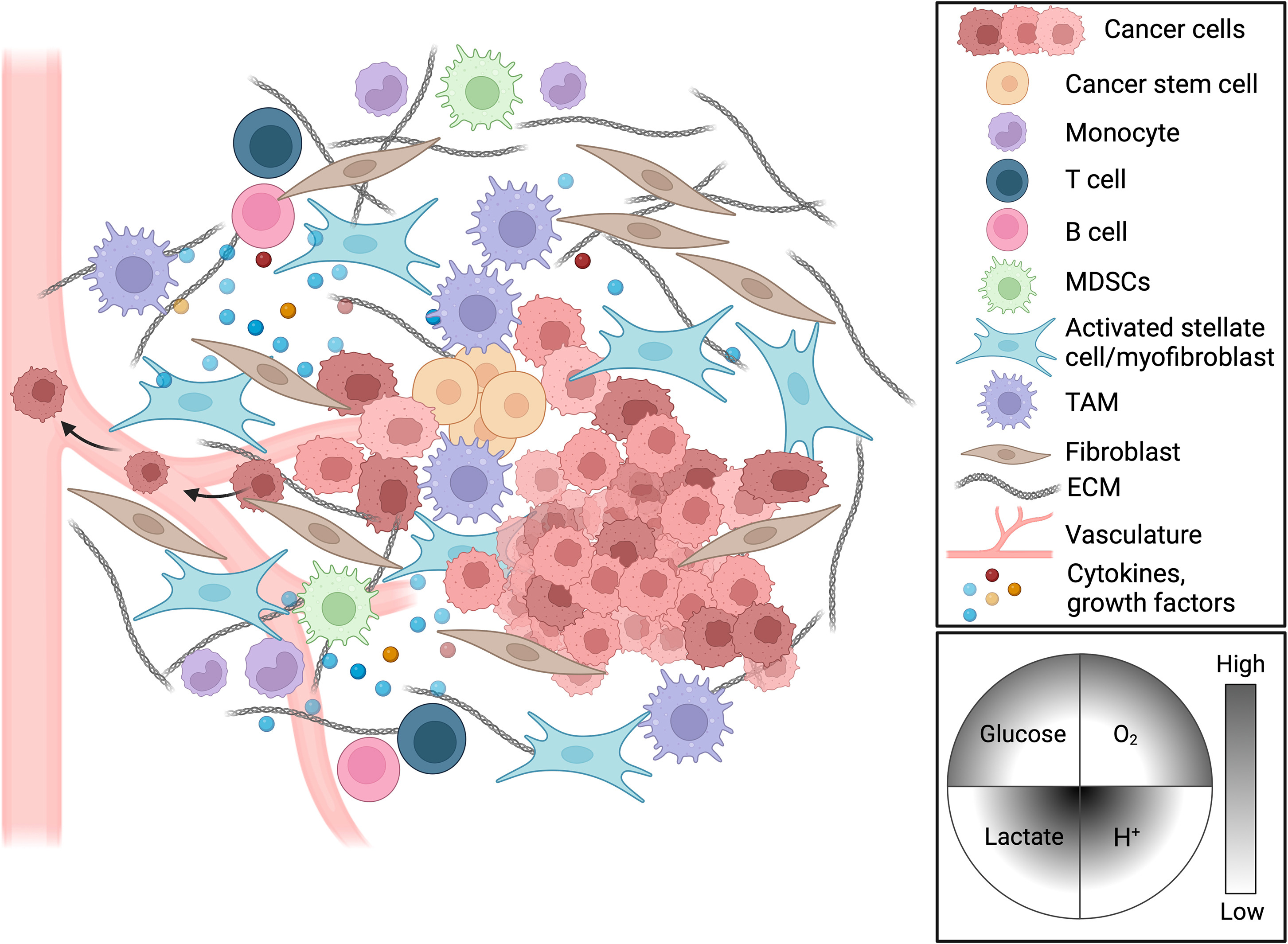
Figure 1: Complex cellular composition of the tumor microenvironment.
Tumor tissue is not just a collection of cancer cells, but also contains various stromal and immune cells such as tumor-associated macrophages (TAM), myeloid-derived suppressor cells (MDSC), T cells, B cells, dendritic cells, and cancer-associated fibroblasts (CAF). Accurate analysis of the composition and functional status of these cell subsets requires efficient tissue dissociation techniques to release cells intact.
Why is tumor tissue dissociation so challenging?
The tissue structure of solid tumors is distinctly different from that of normal tissues, and this "abnormal" structure poses multiple barriers to cell isolation.
Dense Stromal Barrier
Tumor tissues are often accompanied by significant stromal deposition:
- Collagen Remodeling: Cancer-associated fibroblasts (CAF) abundantly secrete type I and III collagen, forming a dense fibrous network
- Stromal Cross-linking: Lysyl oxidase (LOX)-mediated collagen cross-linking increases the mechanical strength of the stroma
- Abnormal Vasculature: Disordered tumor blood vessel structure leads to local hypoxia and necrotic areas, impairing enzyme penetration
Conventional single-enzyme digestion with trypsin or collagenase struggles to address multiple stromal components simultaneously, resulting in incomplete dissociation or excessively long digestion times.
Extreme Heterogeneity of Cell Types
The tumor microenvironment contains dozens of cell types, which vary drastically in their sensitivity to digestion conditions:
- Fragile Lymphocytes: Surface antigens (e.g., CD3, CD4, CD8) are susceptible to proteolytic degradation, and over-digestion causes false negatives
- Robust Tumor Cells: Tight intercellular junctions between epithelial cells require strong digestive power for separation
- Large Macrophages: Prone to rupture during mechanical grinding, releasing RNases and proteases that damage other cells
A single digestion condition cannot balance the survival and antigen integrity of all cell types.
Interference of Immunosuppressive Microenvironment
Immunosuppressive factors (e.g., TGF-β, IL-10) and metabolic waste (e.g., lactic acid) in the tumor microenvironment affect the state of dissociated cells:
- Reduced Cell Viability: Low pH and hypoxic conditions make cells more sensitive to enzymatic digestion
- Surface Antigen Modification: Matrix metalloproteinases (MMP) in the tumor microenvironment may partially cleave cell surface molecules
- Cell Aggregation Tendency: Dissociated cells may re-aggregate, compromising single-cell suspension quality
How do specialized kits solve these challenges?
Tumor Tissue Dissociation Kits achieve the balance between "efficient dissociation" and "cell protection" through three core technical strategies:
Multi-enzyme Synergistic Formulation
Unlike single collagenase, specialized kits employ an optimized combination of multiple enzymes:
- Collagenase: Breaks down collagen fibers, targeting the main component of tumor stroma
- Neutral Protease/Dispase: Cleaves intercellular junction proteins (e.g., E-cadherin) to facilitate epithelial cell separation
- Elastase: Degrades elastic fibers, particularly suitable for tumors rich in elastin such as lung and skin tumors
- DNase: Degrades DNA released by dead cells, reducing cell aggregation and viscosity
The activity ratios of these enzymes are optimized to ensure synergistic action rather than mutual inhibition.
Mild Digestion Conditions
- Temperature Control: Incubation at 37°C, close to physiological temperature, with moderate enzyme activity
- Time Optimization: Standard digestion time of 30-40 minutes to avoid over-digestion
- Mechanical Assistance: Gentle shaker oscillation (80rpm) promotes enzyme penetration and reduces mechanical shear force
Selective Enrichment Strategy
Percoll density gradient centrifugation enables:
- Removal of Cell Debris: Necrotic tissue and undigested stroma sediment to the bottom of the tube
- Enrichment of Viable Cells: Viable cells match the Percoll gradient density, forming a distinct buffy coat
- Isolation of Specific Subsets: Adjusting Percoll concentrations (e.g., 40%/80% double-layer gradient) allows enrichment of lymphocytes or tumor cells
Which experimental scenarios require specialized dissociation kits?
Analysis of Tumor-Infiltrating Lymphocytes (TILs)
This is the core application scenario in tumor immunology research. The composition and phenotypic status of TILs are key indicators for evaluating anti-tumor immune responses and predicting the efficacy of immune checkpoint inhibitors.
Specialized kits ensure:
- Complete Retention of T Cell Subsets: Key markers such as CD3, CD4, CD8, and FoxP3 are not degraded
- Viability of Myeloid Cells: Sustained viability of tumor-associated macrophages (TAM) and MDSCs to support functional assays
- NK Cells and Dendritic Cells: Efficient recovery of rare cell subsets to avoid loss during cumbersome digestion
Single-Cell Sequencing (scRNA-seq)
Single-cell transcriptomic analysis has extremely high requirements for cell quality:
- Cell Viability >90%: Mild conditions of specialized kits reduce cell stress and death
- No Cell Aggregation: DNase treatment minimizes cell clumps and avoids "doublet" artifacts
- Fidelity of Gene Expression: Prevents abnormal expression of heat shock proteins and immediate-early genes induced by digestive stress
- Tumor-Immune Interaction Analysis: Fully preserves the expression of receptors involved in intercellular communication
Establishment of Patient-Derived Organoids (PDO)
The establishment of patient-derived tumor organoids relies on the survival of viable tumor cells:
- Preservation of Stem Cell Activity: Mild enzymatic conditions protect the self-renewal capacity of cancer stem cells
- Removal of Stromal Cells: Percoll purification selectively removes CAFs to obtain relatively pure tumor cells
- Support for Long-Term Culture: High viability of dissociated cells facilitates organoid formation in Matrigel
Recommended Absin Tumor Tissue Dissociation Kits
| Cat. No. | Product Name | Size |
|---|---|---|
| abs50099 | Human Tumor Tissue Dissociation Kit | 10T/25T |
| abs50090 | Rat/Mouse Tumor Tissue Dissociation Kit | 10T/25T |
Conclusion
Tumor Tissue Dissociation Kits represent the evolution of tumor sample preparation technology from "experience-driven" to "standardized workflows". In the era of booming cancer immunotherapy, precise dissection of the cellular composition and functional status of the tumor microenvironment hinges on high-quality sample preparation. From TIL analysis to single-cell sequencing, from organoid establishment to drug screening, specialized dissociation technology is always the first step to obtaining reliable data. Mastering the essence of this technology means establishing a quality advantage at the starting line of cancer research.
Contact Absin
Absin provides antibodies, proteins, ELISA kits, cell culture, detection kits, and other research reagents. If you have any product needs, please contact us.
| Absin Bioscience Inc. worldwide@absin.cn |
 Follow us on Facebook: Absin Bio Follow us on Facebook: Absin Bio |