- Cart 0
- English
How does the Coomassie Brilliant Blue Staining Kit make protein bands clearly visible?
May 08, 2026
Clicks:72
Protein electrophoresis is one of the most fundamental techniques in molecular biology research, and gel staining is a critical step for obtaining experimental results. Among various protein staining methods, Coomassie Brilliant Blue staining has become the most commonly used protein visualization tool in laboratories due to its advantages of simple operation, low cost, and moderate sensitivity. This article will deeply discuss the composition characteristics, staining principle and applications of Coomassie Brilliant Blue Staining Kit in various experiments.
What is Coomassie Brilliant Blue Staining Kit?
Coomassie Brilliant Blue Staining Kit adopts the most classic Coomassie Brilliant Blue staining and destaining methods, using Coomassie Brilliant Blue R250 as the dye. It can be used for routine staining and destaining of protein electrophoresis gels such as SDS-PAGE or native PAGE, or detection of residual proteins on PAGE gels after Western transfer.
The kit usually contains two core components: Coomassie Brilliant Blue Staining Solution (100mL) and Coomassie Brilliant Blue Staining Destaining Solution (500 mL). The staining solution contains Coomassie Brilliant Blue R250 dye, methanol, acetic acid and other components, while the destaining solution contains methanol and acetic acid to remove gel background staining.
Coomassie Brilliant Blue R250 is a triphenylmethane dye. When bound to proteins under acidic conditions, its maximum absorption wavelength shifts from 465 nm (reddish-brown) to 595 nm (blue), with an obvious color change for easy observation.
Why Does Coomassie Brilliant Blue Bind to Proteins?
Hydrophobic interaction is the main mechanism for the binding of Coomassie Brilliant Blue to proteins. Coomassie Brilliant Blue molecules possess a hydrophobic aromatic ring structure, which can interact with hydrophobic regions in protein molecules (such as side chains of aromatic amino acids) via hydrophobic forces.
Electrostatic interaction also participates in the binding process. In acidic staining solution, proteins are positively charged and Coomassie Brilliant Blue dye is negatively charged. The electrostatic attraction between positive and negative charges enhances the binding of the dye to proteins.
Principle of color change: Free Coomassie Brilliant Blue R250 appears reddish-brown (max absorption 465nm), and turns blue (max absorption 595nm) after binding to proteins. This color change makes protein bands clearly visible in the gel.
What Experimental Scenarios Can It Be Applied To?
SDS-PAGE protein electrophoresis is the most basic application of Coomassie Brilliant Blue staining. After electrophoresis separation, protein bands can be stained to intuitively determine the purity, molecular weight and expression level of protein samples. The molecular weight of the target protein can be estimated by comparison with protein Marker; the protein expression levels of different samples can be semi-quantitatively compared through band gray analysis.
Native PAGE electrophoresis is also applicable. Proteins separated under non-denaturing conditions retain their native conformations, and Coomassie Brilliant Blue staining can be used for experiments such as protein complex detection and enzyme activity analysis.
Western Blot transfer efficiency evaluation is another important application. After membrane transfer, staining the residual proteins on the PAGE gel with Coomassie Brilliant Blue can intuitively evaluate whether the transfer is complete. If obvious protein bands remain on the gel, it indicates insufficient transfer; if no protein remains on the gel but signals are present on the membrane, the transfer is successful.
Protein purification process monitoring relies on Coomassie Brilliant Blue staining. In purification steps such as affinity chromatography, ion exchange chromatography and gel filtration, the protein content and purity of each eluted fraction are detected by staining to optimize purification conditions and determine the elution peak of the target protein.
Protein concentration estimation can be performed using Coomassie Brilliant Blue staining. By comparing the band grayness with protein standards of known concentrations, the sample protein concentration can be roughly estimated as a reference for subsequent experiments.
In proteomics analysis, Coomassie Brilliant Blue staining is used for visualization of protein spots on 2D electrophoresis gels, combined with mass spectrometry for protein identification.
How to Operate the Conventional Staining and Destaining Method?
Staining steps:
After electrophoresis, place the gel in an appropriate amount of Coomassie Brilliant Blue Staining Solution to ensure the gel is fully covered. Shake gently on a horizontal shaker or side-to-side shaker and stain at room temperature for 1 hour or longer.
The specific staining time depends on the gel thickness and staining temperature. Prolong the staining time appropriately for thicker gels or lower temperatures; shorten the time for thinner gels or higher temperatures. Generally, sufficient staining is achieved when the gel color is very close to that of the staining solution and the gel is barely visible in the solution.
The staining solution can be recycled and reused at least 2-3 times to reduce experimental costs.
Destaining steps:
Pour out the staining solution and add an appropriate amount of Coomassie Brilliant Blue Destaining Solution to fully cover the gel. Shake gently on a horizontal shaker or side-to-side shaker and destain at room temperature for 4-24 hours. Replace the destaining solution 2-4 times during the process until the blue background is almost completely removed and the protein band staining meets expectations.
Usually, protein bands appear after 1-2 hours of destaining, but complete background removal requires a longer time. Excessive destaining time will lighten the color of protein bands, so a balance between background clarity and band intensity is required.
Gel preservation:
After destaining, the gel can be stored in water for subsequent photography and other operations. Gels stored in water will swell; to prevent swelling, store the gel in water containing 20% glycerol. Dry gels can be prepared for long-term storage.
How to Operate the Rapid Staining and Destaining Method?
For time-sensitive experiments, the rapid staining and destaining method can be adopted:
Rapid staining:
After electrophoresis, place the gel in an appropriate amount of Coomassie Brilliant Blue Staining Solution, heat in a microwave until near boiling or just boiling, then stop heating immediately. Gels above 10% are usually tough and not easy to break during boiling; for gels below 10%, boiling should be avoided as much as possible to prevent gel fragmentation. Then shake on a room-temperature shaker for 5-10 minutes while the staining solution is still hot.
Rapid destaining:
Pour out the staining solution and add an appropriate amount of Coomassie Brilliant Blue Destaining Solution, heat in a microwave until near boiling or just boiling, then stop heating immediately. Then shake on a shaker for 5-10 minutes while the destaining solution is still hot. Clear protein bands can usually be observed at this point.
Replace with fresh destaining solution and repeat the heating and shaking steps until the blue background is almost completely removed and the protein band staining meets expectations.
The rapid method can shorten the entire staining and destaining process to 1-2 hours, but attention should be paid to controlling the heating temperature and time to avoid gel breakage or protein band diffusion.
What Are the Key Precautions for Use?
- Staining time optimization. Insufficient staining time results in faint protein bands and reduced sensitivity; excessive staining increases background staining and prolongs subsequent destaining time. It is recommended to optimize according to gel thickness (0.75mm, 1.0mm or 1.5mm) and temperature conditions.
- Destaining solution replacement frequency. The destaining solution gradually becomes saturated with dye during destaining. Timely replacement of fresh destaining solution accelerates background clearance. Generally, replace every 4-6 hours until the background is clear.
- Staining solution recycling. The staining solution can be recycled and reused at least 2-3 times, but staining efficiency gradually decreases with increased usage times. It is recommended to record the usage times and replace with new staining solution in a timely manner.
- Safety protection. Coomassie Brilliant Blue dye and methanol in the destaining solution are harmful to the human body. Proper protection is required during operation, such as wearing lab coats and gloves, and performing operations in a fume hood.
- Gel thickness consideration. Prolong staining and destaining time appropriately for thick gels (1.5mm) and shorten for thin gels (0.75mm). In the rapid method, boiling of low-concentration gels (<10%) should be avoided to prevent fragmentation.
- Temperature control. Staining and destaining at room temperature are standard conditions. Low temperatures slow down the staining and destaining rate, while high temperatures may accelerate methanol volatilization or cause gel deformation.
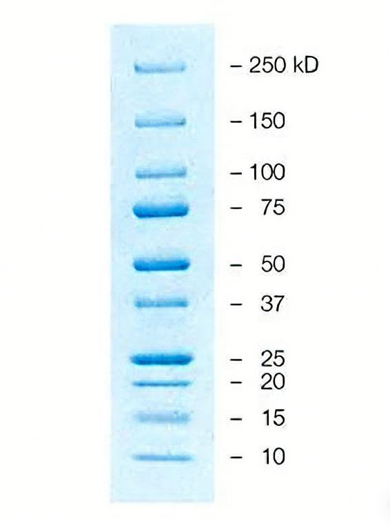
SDS-PAGE Protein Electrophoresis Staining Result
Conclusion
As a classic supporting reagent for protein electrophoresis, Coomassie Brilliant Blue Staining Kit occupies an important position in life science research due to its advantages of simple operation, low cost and reliable results. From routine protein expression detection to complex proteomics analysis, from teaching laboratories to high-throughput screening platforms, Coomassie Brilliant Blue staining is an indispensable protein visualization tool. Mastering both conventional and rapid staining/destaining methods, optimizing operating conditions according to experimental needs, and paying attention to safety protection and reagent recycling will ensure clear and accurate protein band results. With the development of digital imaging technology, the quantitative analysis capability of Coomassie Brilliant Blue staining will be further enhanced, providing more accurate data support for protein research.
Absin Coomassie Brilliant Blue Staining Kit Recommendation:
| Cat. No. | Product Name | Size |
|---|---|---|
| abs964 | Coomassie Brilliant Blue Staining Kit (Conventional) | 1kit |
Contact Absin
Absin provides antibodies, proteins, ELISA kits, cell culture, detection kits, and other research reagents. If you have any product needs, please contact us.
| Absin Bioscience Inc. worldwide@absin.cn |
 Follow us on Facebook: Absin Bio Follow us on Facebook: Absin Bio |