- Cart 0
- English
How does the Mitochondrial Respiratory Chain Complex Activity Assay Kit reveal the mysteries of cellular energy metabolism?
May 07, 2026
Clicks:72
As the powerhouse of the cell, the functional status of the mitochondrial respiratory chain directly reflects cellular metabolic health and physiological function. Mitochondrial respiratory chain complexes (Complex I-V) are the core components of the oxidative phosphorylation system. Dysfunction of any complex may lead to energy metabolism disorders, increased reactive oxygen species production, and is further associated with the occurrence and development of various diseases. Mitochondrial Respiratory Chain Complex Activity Assay Kits provide reliable technical tools for accurately evaluating the activity of these key enzymes.
What is Mitochondrial Respiratory Chain Complex?
The mitochondrial respiratory chain consists of five protein complexes (Complex I-V) located on the inner mitochondrial membrane, which are key structures for cellular aerobic respiration and ATP production.
Complex I (NADH-CoQ reductase or NADH dehydrogenase) is the largest protein complex in the inner mitochondrial membrane. It catalyzes electron transfer from NADH to CoQ and is the main site of superoxide anion (O₂⁻) production in the respiratory electron transport chain (ETC). Measuring its activity can reflect both the ETC status and reactive oxygen species (ROS) generation.
Complex II (Succinate-coenzyme Q reductase) catalyzes the oxidation of succinate to fumarate, while the prosthetic group FAD is reduced to FADH₂, which further reduces oxidized coenzyme Q to reduced coenzyme Q, serving as a branch of the respiratory electron transport chain.
Complex III (CoQ-cytochrome c reductase) transfers hydrogen from reduced CoQ to cytochrome c to generate reduced cytochrome c, and is a common component of both the main and branch pathways of the mitochondrial respiratory electron transport chain.
Complex IV (Cytochrome c oxidase) catalyzes the oxidation of reduced cytochrome c and ultimately transfers electrons to oxygen to form water, also a common component of the main and branch pathways of the mitochondrial electron transport chain.
Complex V (F₁F₀-ATP synthase) utilizes the proton electrochemical gradient generated by the respiratory chain to catalyze ATP synthesis and can also hydrolyze ATP in the reverse process. It is a key enzyme for ATP synthesis via oxidative phosphorylation in mitochondria and photophosphorylation in chloroplasts.
Why Detect Complex Activity?
Disease mechanism research requires precise assessment of complex function. Mitochondrial dysfunction is closely related to various diseases such as neurodegenerative diseases (e.g., Parkinson's disease, Alzheimer's disease), cardiovascular diseases, metabolic syndrome, and tumors. Detecting activity changes of specific complexes can reveal the molecular mechanisms of disease pathogenesis.
Drug screening and toxicity evaluation rely on complex activity detection. Many drugs (e.g., anti-tumor drugs, antibiotics) target the mitochondrial respiratory chain, and complex activity detection can evaluate their mechanism of action and mitochondrial toxicity.
Cellular metabolic status assessment can be reflected by complex activity. Metabolic reprogramming of cells under hypoxia, nutrient deficiency, or stress is often accompanied by changes in respiratory chain function.
Plant physiological research also requires complex activity data. Although plant mitochondrial respiratory chains differ from animals, the basic functions of Complex I-V are conserved, and this kit is also suitable for plant tissue and cell samples.
What is the Detection Principle?
The detection principles of different complexes are based on their specific enzymatic reactions and substrate-product characteristics:
Complex I Detection Principle: Mitochondrial respiratory chain Complex I catalyzes the dehydrogenation of NADH to NAD⁺. The enzyme activity is calculated by measuring the oxidation rate of NADH at 340 nm. NADH has a characteristic absorption peak at 340 nm, and its oxidation leads to a decrease in absorbance.
Complex II Detection Principle: Reduced coenzyme Q, the catalytic product of Complex II, further reduces 2,6-dichloroindophenol, which has a characteristic absorption peak at 605 nm. The enzyme activity is calculated by detecting the reduction rate of 2,6-dichloroindophenol.
Complex III Detection Principle: Complex III transfers hydrogen from reduced CoQ to cytochrome c to generate reduced cytochrome c. Unlike oxidized cytochrome c, reduced cytochrome c has a characteristic light absorption at 550 nm, so the increase rate of absorbance at 550 nm reflects Complex III activity.
Complex IV Detection Principle: Reduced cytochrome c has a characteristic light absorption at 550 nm. Complex IV catalyzes the conversion of reduced cytochrome c to oxidized cytochrome c, so the decrease rate of absorbance at 550 nm reflects Complex IV activity.
Complex V Detection Principle: Complex V hydrolyzes ATP to produce ADP and Pi. The activity is determined by measuring the increase rate of Pi.
What Experimental Scenarios Can Be Applied?
Neurodegenerative disease research: Complex I dysfunction is a key pathological feature of Parkinson's disease. Detecting Complex I activity in patient-derived cells or animal models can evaluate disease progression and therapeutic efficacy.
Tumor metabolism research: Focuses on the relationship between the Warburg effect and respiratory chain function. Tumor cells often exhibit enhanced glycolysis and weakened oxidative phosphorylation, and complex activity detection helps reveal metabolic reprogramming mechanisms.
Drug mitochondrial toxicity screening: Requires systematic evaluation of drug effects on each complex. Certain anti-tumor drugs (e.g., anthracyclines) and antibiotics (e.g., streptomycin) have mitochondrial toxicity, and complex activity detection is an important safety evaluation tool.
Exercise physiology research: Mitochondrial function is a key determinant of muscle endurance. The effects of training interventions on mitochondrial content and complex activity can be quantitatively assessed with this kit.
Aging research: Focuses on age-related changes in mitochondrial function. Decreased complex activity is an important marker of cellular senescence, and detecting complex activity in samples of different ages helps reveal aging mechanisms.
Plant stress physiology research: Changes in plant mitochondrial respiratory chain function under drought, salt stress, low temperature and other adverse conditions can be evaluated by complex activity detection.
How to Prepare Samples and Perform Detection Correctly?
Mitochondria Extraction Procedure:
- Accurately weigh 0.1 g of tissue or collect 5 million cells, add 1 mL of Reagent I and 10 μL of Reagent III, and homogenize in an ice bath using a homogenizer or mortar.
- Centrifuge the homogenate at 600 g for 5 min at 4°C, collect the supernatant into a new centrifuge tube, and discard the pellet.
- Centrifuge the supernatant again at 11,000 g for 10 min at 4°C; the pellet is the extracted mitochondria. Fresh samples are recommended to ensure enzyme activity.
Complex I Detection Steps:
- Pre-warm the microplate reader or UV spectrophotometer for more than 30 min and adjust the wavelength to 340 nm.
- Add 10 μL of sample, 200 μL of working solution, and 15 μL of Working Reagent VI sequentially to a 96-well UV plate or micro quartz cuvette. Mix thoroughly, then immediately read the initial absorbance A₁ at 0 min and absorbance A₂ after 2 min at 340 nm, and calculate ΔA = A₁ - A₂.
Complex IV Detection Steps:
- Pre-warm the microplate reader and visible spectrophotometer for more than 30 min and adjust the wavelength to 550 nm.
- Add 200 μL of working solution and 10 μL of sample sequentially to a 96-well plate or micro glass cuvette. Mix thoroughly, then immediately read the initial absorbance A₁ at 0 min and absorbance A₂ after 1 min at 550 nm, and calculate ΔA = A₁ - A₂.
Result Calculation:
Calculated based on sample fresh weight. One enzyme activity unit is defined as 1 nmol of substrate consumed (or product produced) per minute per gram of tissue in the reaction system. The kit provides detailed calculation formulas, including derived formulas and simplified formulas, which are completely equivalent.
What are the Key Precautions During Use?
- Sample freshness is critical. Fresh samples are recommended to ensure enzyme activity. If not used immediately, store intact cells or aliquoted tissues at -80°C and use within one month.
- Preliminary experiments are essential. To ensure accuracy, perform a preliminary experiment with 1-2 samples first. If the absorbance or ΔA is too high, dilute the sample with the corresponding reagent before measurement; if ΔA is too small, increase the sample volume to improve detection values; negative ΔA indicates the absence or degradation of the target complex in the sample.
- Reaction time control. This kit is based on enzyme kinetics with fast reactions; reverse reactions may occur after equilibrium. Limit sample groups to 2-3, pre-warm instruments, and perform loading next to the microplate reader for immediate measurement after mixing.
- Strict temperature control. Incubate mammalian samples at 37°C and other species at 25°C. Ensure all components and equipment reach the appropriate temperature before starting the experiment.
- Avoid cross-contamination. Do not mix components from different batch numbers or manufacturers; avoid bubbles when mixing or reconstituting reagents; change pipette tips frequently to prevent cross-contamination.
- Safety protection. Some reagents are toxic or corrosive. Wear lab coats and disposable gloves during operation.
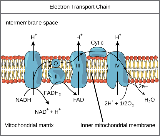 Schematic diagram of mitochondrial electron transport chain
Conclusion
Mitochondrial Respiratory Chain Complex Activity Assay Kits provide accurate and reliable quantitative tools for cellular energy metabolism research. They play important roles in various fields, from basic life science research to clinical disease mechanism exploration, from drug development to environmental toxicology evaluation. Correctly mastering the key technical points of mitochondrial extraction, enzyme activity detection, and result calculation, and strictly controlling experimental conditions and sample quality will ensure accurate and repeatable experimental data. With the advancement of mitochondrial biology research and precision medicine, mitochondrial function assessment technology will show broader application prospects in disease diagnosis, treatment monitoring, and drug development.
Recommended Absin Mitochondrial Respiratory Chain Complex Activity Assay Kits:
| Cat. No. | Product Name | Size |
|---|---|---|
| abs580238 | Micro Mitochondrial Complex I Activity Assay Kit | 96T |
| abs580239 | Micro Mitochondrial Complex II Activity Assay Kit | 96T |
| abs580240 | Micro Mitochondrial Complex III Activity Assay Kit | 96T |
| abs580241 | Micro Mitochondrial Complex IV Activity Assay Kit | 96T |
| abs580242 | Micro Mitochondrial Complex V Activity Assay Kit | 96T |
Contact Absin
Absin provides antibodies, proteins, ELISA kits, cell culture, detection kits, and other research reagents. If you have any product needs, please contact us.
| Absin Bioscience Inc. worldwide@absin.cn |
 Follow us on Facebook: Absin Bio Follow us on Facebook: Absin Bio |