- Cart 0
- English
How to rapidly detect cell viability using trypan blue staining solution?
May 07, 2026
Clicks:72
Cell viability detection is a fundamental step in cell culture experiments, which directly affects the reliability and repeatability of experimental data. Among numerous detection methods, Trypan Blue Staining has become the most commonly used technique for evaluating cell viability in laboratories due to its advantages of simple operation, low cost and intuitive results. This article will deeply discuss the working principle, application scenarios and key operational points of Trypan Blue Stain.
What is Trypan Blue Stain?
Trypan Blue, also known as Direct Blue 3B, is a vital cell dye commonly used to detect cell membrane integrity and cell viability. As an azo compound, it has a large molecular weight and cannot penetrate intact cell membranes.
Trypan Blue Stain is usually supplied as a 0.4% aqueous solution and needs to be diluted to a working concentration (0.04%-0.4%) with PBS or normal saline before use. Its staining principle is based on the difference in cell membrane permeability: cells with lost viability or damaged membrane integrity have impaired membrane barrier function, allowing Trypan Blue to enter the cells and bind to intracellular proteins, staining the cells blue; in contrast, normal living cells with intact membranes can exclude Trypan Blue and remain unstained. Therefore, the loss of cell membrane integrity is generally regarded as a marker of cell death.
Why Can Trypan Blue Rapidly Distinguish Live and Dead Cells?
Differential cell membrane permeability is the core mechanism of Trypan Blue staining. The cell membrane of living cells possesses selective permeability, and Trypan Blue molecules cannot cross the intact lipid bilayer due to their negative charge and large molecular weight (approximately 960 Da). In dead cells, the membrane integrity is disrupted, forming pores or complete disintegration, allowing free entry of Trypan Blue into the cytoplasm.
Obvious optical contrast enables intuitive result interpretation. After staining, dead cells are blue, swollen and non-lustrous, while living cells remain unstained with normal morphology and luster. This distinct visual difference allows rapid discrimination between live and dead cells under a standard light microscope without specialized equipment.
Rapid staining speed is another remarkable advantage. Trypan Blue staining can be completed in 3-5 minutes at room temperature. Accurate quantification of cell viability can be achieved by direct counting under a microscope or post-imaging counting, with the entire process finished within 10 minutes.
What Experimental Scenarios Require Trypan Blue Staining?
Routine quality control of cell culture is the most basic application of Trypan Blue staining. Before cell passage, cryopreservation or experimental treatment, it is necessary to assess cell status and health. Calculating viability via Trypan Blue staining determines whether cells are suitable for further culture or subsequent experiments. Generally, cells with viability below 90% are not recommended for precise experiments.
Drug toxicity screening widely employs Trypan Blue staining. In experiments such as anti-tumor drug development and compound toxicity evaluation, the cytotoxic effects of drugs or compounds can be preliminarily assessed by comparing cell viability between treatment and control groups. This method is suitable for preliminary evaluation in high-throughput screening.
Evaluation of cell cryopreservation and resuscitation efficiency relies on Trypan Blue staining. DMSO in cryopreservation medium and low-temperature processes may damage cells. Detecting viability after resuscitation using Trypan Blue helps optimize cryopreservation conditions (e.g., cooling rate, cryopreservation medium formulation) and resuscitation protocols.
Immune cell function research has special applications for Trypan Blue. Macrophages can phagocytose Trypan Blue, making it a vital stain for macrophages. The phagocytic activity of macrophages can be evaluated by observing the proportion of Trypan Blue-positive cells.
Verification of cell separation and purification efficiency also requires Trypan Blue staining. After flow cytometry sorting, magnetic bead sorting or density gradient centrifugation, the purity and viability of isolated cells are assessed by Trypan Blue staining to ensure the reliability of subsequent experiments.
Apoptosis detection auxiliary method can also use Trypan Blue. Note that apoptotic bodies exhibit Trypan Blue exclusion, so this staining cannot distinguish apoptotic cells from living cells, only necrotic cells from living cells. For apoptosis detection, comprehensive judgment is required in combination with methods such as Annexin V/PI double staining.
How to Perform Trypan Blue Staining Correctly?
Sample Preparation:
Collect cells; adherent cells may require digestion with trypsin and/or EDTA. Centrifuge at 1000 r/min for 5 minutes, discard the supernatant, prepare a single-cell suspension, and dilute appropriately to achieve a suitable cell density for counting (typically 1×10⁵-1×10⁶ cells/mL).
Staining Operation:
Add 0.4% Trypan Blue solution to the cell suspension and mix thoroughly at an appropriate ratio (final Trypan Blue concentration 0.04%-0.4%). Stain at room temperature for 3 minutes; the staining time can be extended appropriately but should not exceed 10 minutes. Excessive staining time causes living cells to gradually accumulate the dye and become stained, leading to biased detection results.
Microscopic Counting:
Aspirate a small amount of stained cells and count using a hemocytometer. Under the microscope: dead cells are blue, swollen and non-lustrous; living cells are unstained with normal morphology and luster. At least 500 cells should be counted to ensure statistical accuracy.
Viability Calculation:
Cell Viability (%) = Total Number of Live Cells / (Total Number of Live Cells + Total Number of Dead Cells) × 100%
What are the Key Precautions During Operation?
- Strict control of staining time. Excessive staining time causes living cells to accumulate the dye and produce false-positive results, leading to detection bias. The recommended staining time is 3-5 minutes, with a maximum of 10 minutes.
- Sufficient cell count. For accurate quantification, at least 500 cells should be counted per sample. Insufficient cell number increases statistical errors and reduces result reliability.
- Crystal handling. Crystals may precipitate in low-temperature environments. Rewarm the reagent in a 37°C water bath before use until crystals dissolve completely. Repeated freeze-thaw cycles accelerate crystal formation; aliquot storage is recommended.
- Concentration optimization. The working concentration of Trypan Blue is generally 0.04%-0.4%, which can be optimized according to cell type and experimental purpose. Low concentration may result in insufficient staining of dead cells, while high concentration increases background staining.
- Single-cell suspension preparation. Ensure complete cell dispersion to avoid clumps affecting counting accuracy. For easily aggregating cell types, increase pipetting times or use EDTA-containing buffer.
- Microscope adjustment. A standard light microscope with 10× or 20× objective lenses is recommended. Adjust the aperture and contrast appropriately to maximize the contrast between blue dead cells and transparent living cells for accurate counting.
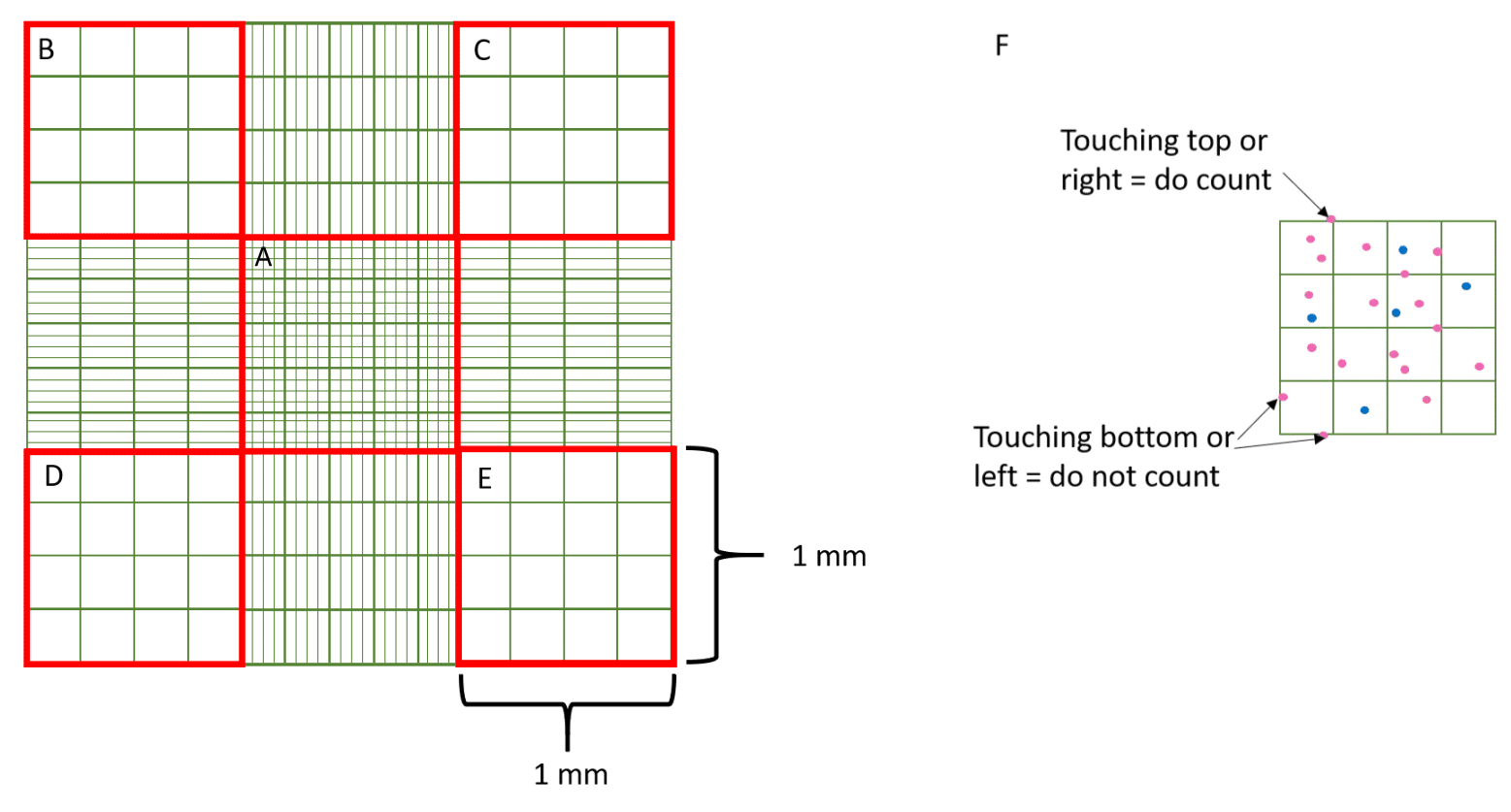 Schematic diagram of cell counting with hemocytometer
Conclusion
As a classic tool for cell viability detection, Trypan Blue Stain occupies an important position in cell biology research due to its simplicity, rapidity and cost-effectiveness. From daily cell culture quality control to drug screening, from cryopreservation efficiency evaluation to immune cell function research, Trypan Blue staining provides reliable cell viability data. Mastering key points such as staining time, cell number and counting methods ensures the accuracy and repeatability of detection results. With the popularization of automated cell counters, Trypan Blue Staining is gradually integrated with digital technology, providing more efficient and precise solutions for cell viability detection.
Absin Trypan Blue Stain Recommendation:
| Cat. No. | Product Name | Size |
|---|---|---|
| abs47047622 | Trypan Blue Stain (0.4%) | 50mL/100mL |
Contact Absin
Absin provides antibodies, proteins, ELISA kits, cell culture, detection kits, and other research reagents. If you have any product needs, please contact us.
| Absin Bioscience Inc. worldwide@absin.cn |
 Follow us on Facebook: Absin Bio Follow us on Facebook: Absin Bio |