- Cart 0
- English
How does the azure-eosin composite dye achieve accurate identification of blood cells?
April 28, 2026
Clicks:72
In the fields of hematology and clinical diagnosis, the staining quality of blood smears directly determines the accuracy of white blood cell differential counts. Giemsa Stain, a composite staining system composed of the basic dye Azure and the acidic dye Eosin, enables distinct coloration of different cellular components through pH-dependent charge interactions, thereby clearly differentiating various blood cell types such as neutrophils, lymphocytes, and eosinophils. As an improved version of Wright staining, Giemsa staining has become a classic method for blood smear staining, bone marrow smear analysis, and pathogen detection due to its advantages of vivid staining, clear cellular structure, and long preservation time.
How Does the Azure-Eosin Composite Dye Achieve Differential Staining of Cellular Components?
The staining principle of Giemsa Stain is based on the ampholyte properties of different chemical components in cells and the selective binding of dyes:
Binding of Basic Dye Azure: DNA in the cell nucleus and lymphocyte cytoplasm are rich in phosphate groups, which are acidic (negatively charged) at physiological pH. As a basic dye, Azure carries a positive charge and binds to acidic groups via electrostatic attraction, staining the nucleus and lymphocyte cytoplasm purple-blue or dark blue.
Binding of Acidic Dye Eosin: Eosinophilic granules are mainly composed of basic proteins (e.g., histones, protamines), which are basic (positively charged) under staining conditions. Eosin, as an acidic dye, carries a negative charge and binds to basic granules, staining the specific granules of eosinophils a bright pink or red.
Double Staining of Neutrophilic Granules: Neutrophilic granules are isoelectric and can bind to both Azure and Eosin, finally presenting light purple or pale purplish red, which is the origin of the name "neutrophil".
General Cytoplasmic Staining: The cytoplasm of most cells, due to its high protein content, usually presents a pale pink color after eosin staining, forming a sharp contrast with the deeply stained nucleus.
How Does pH Act as an Invisible Regulator of Staining Success?
Giemsa staining is extremely sensitive to hydrogen ion concentration, and slight changes in pH can lead to staining hue shifts and affect cell identification:
Acidic Environment (pH < 6.8): Proteins gain more positive charges and tend to bind with the acidic dye Eosin, resulting in reddish staining. At this time, the nucleus may appear reddish purple instead of standard purple-blue, and lymphocyte cytoplasm may be over-stained red, leading to difficulties in cell type identification.
Alkaline Environment (pH > 7.2): Proteins gain more negative charges and tend to bind with the basic dye Azure, resulting in bluish staining. The nucleus may be over-stained blue, and eosinophilic granules may lose their typical bright red color and appear blue-gray, also affecting the accuracy of classification.
Optimal pH Range: It is generally recommended to control the pH of the working staining solution between 6.8 and 7.2. At this pH, the nucleus shows clear purple-blue, eosinophilic granules show bright red, and neutrophilic granules show light purple, with distinct contrast between components for easy identification.
Importance of Buffer Solution: The buffer solution provided with the kit (usually pH 6.8-7.0) is not only used to dilute the stock solution but also plays a role in stabilizing the pH of the staining environment. Using clean glass slides and rinse water close to neutral (pH 6.5-7.5) is key to ensuring staining consistency.
Which Experimental Scenarios Best Reflect the Diagnostic Value of Giemsa Staining?
Peripheral Blood Smear White Blood Cell Differentiation
This is the most classic application scenario. Giemsa staining can clearly distinguish:
- - Neutrophils: Nucleus is purple-blue and lobed; cytoplasm is pale pink with light purple neutrophilic granules
- - Lymphocytes: Nucleus is dark purple-blue, round or reniform; cytoplasm is sky blue and scanty
- - Eosinophils: Nucleus is purple-blue; cytoplasm contains coarse bright red eosinophilic granules
- - Basophils: Nucleus is purple-blue, often masked by granules; cytoplasm contains coarse purplish-black basophilic granules
- - Monocytes: Nucleus is purple-blue, reniform or horseshoe-shaped; cytoplasm is grayish-blue with fine dust-like granules
Bone Marrow Cytology Examination
In the diagnosis of hematological diseases, Giemsa staining is used to evaluate the maturity, proportional distribution, and morphological abnormalities of bone marrow cells, serving as an important basis for the diagnosis of leukemia, myelodysplastic syndrome, and other diseases.
Blood Parasite Detection
Giemsa staining is the gold-standard method for detecting blood parasites such as Plasmodium, Trypanosoma, Rickettsia, and Mycoplasma. All developmental stages of Plasmodium in erythrocytes (ring forms, trophozoites, schizonts, gametocytes) show clear blue-purple cytoplasm and red nuclear material after Giemsa staining, facilitating identification and species differentiation.
Cell Culture and Chromosome Analysis
In some cell culture experiments, Giemsa staining is used to observe cell morphology and growth status. In addition, Giemsa banding (G-banding) is an important method for chromosome karyotype analysis, identifying chromosomal abnormalities through the light and dark banding patterns of Giemsa staining.
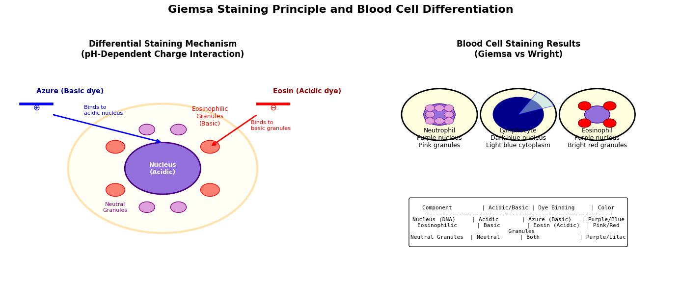
Figure: Schematic diagram of Giemsa staining principle and blood cell differentiation. The left panel shows the pH-dependent charge interaction mechanism: the basic dye Azure (positive charge) binds to acidic nuclei (negative charge) to produce purple-blue; the acidic dye Eosin (negative charge) binds to basic eosinophilic granules (positive charge) to produce red; neutrophilic granules bind to both dyes to produce light purple. The right panel shows staining results: neutrophils (purple nucleus with pink granules), lymphocytes (dark blue nucleus with light blue cytoplasm), eosinophils (purple nucleus with bright red granules).
How to Avoid Common Operational Pitfalls for Ideal Staining Results?
Working Solution Preparation
- Dilution Ratio: Dilute the Giemsa stock solution 20-fold with buffer (e.g., 5 mL stock solution + 95 mL buffer) to obtain the working solution. The stock solution is highly concentrated, and direct use will result in overly deep and uneven staining.
- Prepare Fresh: The working staining solution is unstable and is recommended to be prepared fresh before use, with a storage time not exceeding 48 hours. Old staining solution leads to poor staining effect and dull cell coloration due to oxidative decomposition of dyes.
Smear Preparation and Fixation
- Smear Quality: Blood smears should be uniform in thickness, showing a "tongue-shaped" distribution with clear head, body, and tail regions. Overly thick smears cause uneven staining, while overly thin smears have sparse cell distribution.
- Fixation Conditions: Fix with methanol or ethanol for 10 minutes and air-dry. Insufficient fixation causes cell detachment, while excessive fixation may affect staining affinity.
Staining Time Control
- Standard Time: Generally stain for 45 minutes, but adjust according to cell density and smear thickness. It is best to observe under a microscope during staining, and terminate when the nucleus and granules are clearly stained.
- Over-Staining Treatment: If staining is too deep, perform instantaneous decolorization with 95% ethanol (a few seconds) and immediately wash with water to terminate. Excessive decolorization leads to overly light staining.
- Under-Staining Remedy: If staining is too light, restaining can be performed, but the effect is usually less vivid than one-time staining.
Washing Techniques
- Water Flow Control: The water flow during rinsing should not be too strong to avoid cell detachment; nor too weak to avoid dye residue adhesion. The optimal flow is fine and continuous running water, rinsing at a 30-degree angle.
- Washing Time: Wash thoroughly until the smear is pink or light purple with no dye residue flowing. Insufficient washing causes a deep background, while excessive washing may lead to fading.
Microscopic Examination and Preservation
- Drying Conditions: Examine microscopically after air drying; avoid heat drying which causes cell shrinkage.
- Mounting for Preservation: For long-term preservation, mount with neutral balsam to avoid fading and drying.
How to Weigh the Similarities and Differences with Wright Staining for Selection?
Giemsa staining and Wright Stain share similar principles, both using Azure-Eosin composite dyes, but have the following differences:
- Staining Speed: Wright staining is fast (a few minutes), suitable for emergency rapid screening; Giemsa staining is slower (45-60 minutes) but provides more vivid staining and clearer details.
- Staining Effect: Giemsa staining provides better staining for nuclei and parasites, making it the first choice for blood parasite examination; Wright staining provides better cytoplasmic staining with more natural overall cell morphology.
- Buffer Requirement: Giemsa staining is more sensitive to pH and requires a matching buffer; Wright staining is relatively tolerant.
- Application Scenarios: Wright staining can be used for routine blood screening, while Giemsa staining is recommended for parasite detection and fine cytological examination. The two can also be used in combination (Wright-Giemsa composite staining) to complement each other.
Conclusion
With its classic Azure-Eosin combination and pH-sensitive differential staining characteristics, the Giemsa Stain Kit has become an indispensable tool in hematological diagnosis. From peripheral blood leukocyte differentiation to bone marrow cell morphological analysis, from Plasmodium detection to chromosome banding, Giemsa staining provides reliable morphological evidence for clinical diagnosis and basic research with its stable staining effect and clear cell identification. Mastering key technical points such as pH control, fresh preparation, and fine washing is crucial to achieving ideal staining results and ensuring diagnostic accuracy.
Absin Giemsa Stain Kit Recommendation:
Contact Absin
Absin provides antibodies, proteins, ELISA kits, cell culture, detection kits, and other research reagents. If you have any product needs, please contact us.
| Absin Bioscience Inc. worldwide@absin.cn |
 Follow us on Facebook: Absin Bio Follow us on Facebook: Absin Bio |