- Cart 0
- English
Why can Western Blot membrane regeneration technology greatly improve the data output efficiency of precious samples?
April 24, 2026
Clicks:73
In Western Blot experiments, completing a full immunodetection workflow—from protein electrophoresis and membrane transfer to antibody incubation and chemiluminescent development—typically takes 1–2 days and consumes valuable samples. However, researchers often need to detect both housekeeping proteins (e.g., GAPDH, β-actin) and target proteins in the same sample, or optimize dilution ratios and incubation conditions for different antibodies. Primary and Secondary Antibody Stripping Buffer, also known as Western Antibody Elution Buffer or Membrane Regeneration Buffer, efficiently removes bound primary and secondary antibodies without affecting the immobilized proteins on the membrane, allowing the same blot to be reused 2–3 times without repeating electrophoresis and transfer. This technique not only drastically saves samples and time but also eliminates errors caused by reloading, significantly enhancing the comparability of experimental data.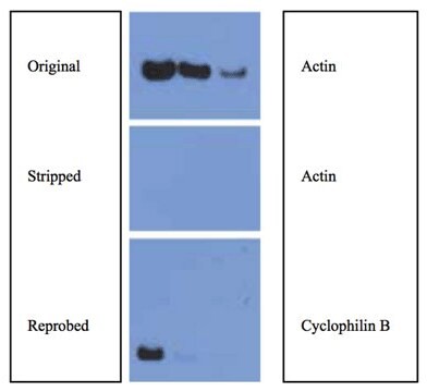
What are the differences between stripping buffers of different pH?
Based on the chemical properties of antibody-antigen interactions, commercially available primary and secondary antibody stripping buffers are mainly classified into four types: neutral, mild alkaline, strong alkaline, and acidic, each suitable for different experimental scenarios and membrane types:
Neutral/Mild Stripping Buffer:
Uses a gentle washing formula to dissociate antibody-antigen interactions via denaturation or competitive binding without damaging target proteins on the membrane. Suitable for most routine Western Blot experiments with minimal protein damage, allowing the membrane to be reused 2–3 times. Especially ideal for subsequent detection of low-abundance target proteins.
Mild Alkaline Stripping Buffer (pH≈10.0):
Mainly disrupts electrostatic interactions and hydrogen bonds between antibodies and antigens under alkaline conditions. This mild alkaline environment is highly compatible with PVDF membranes, effectively stripping primary and secondary antibodies while maintaining protein integrity. Suitable for antibody elution after ECL chemiluminescent detection but not for membrane regeneration after chromogenic detection (e.g., DAB, NBT/BCIP).
Strong Alkaline Stripping Buffer (pH≈11.8):
Provides a stronger alkaline environment for more thorough disruption of antibody-antigen complexes and higher stripping efficiency. Also particularly suitable for PVDF membrane regeneration, with relatively weaker effects on nitrocellulose (NC) membranes. Due to strong alkalinity, it is slightly corrosive; wear gloves and protective equipment during operation.
Acidic Stripping Buffer (pH≈2.0):
Denatures and inactivates antibody proteins under strongly acidic conditions, causing them to detach from the membrane. Acidic formulations achieve complete antibody stripping, suitable for high-affinity antibodies that are difficult to remove or membranes reused multiple times. Note that strong acidic conditions may affect some acid-sensitive proteins; evaluate target protein stability beforehand.
Which types of blotting membranes are best for regeneration?
PVDF Membrane (Polyvinylidene Fluoride):
PVDF membranes exhibit excellent mechanical strength and chemical stability, tolerating stripping buffers of all pH ranges, making them the top choice for membrane regeneration. Their high protein binding capacity and acid-base resistance allow the same PVDF membrane to be reused 2–3 times while maintaining good protein retention.
Nitrocellulose Membrane (NC Membrane):
Although NC membranes can undergo regeneration, the performance is generally inferior to PVDF membranes. NC membranes may partially dissolve or become brittle under strong alkaline conditions, with limited reuse cycles (typically 1–2 times). Therefore, PVDF membranes are preferred if multiple antibody stripping steps are expected.
How to establish a standardized membrane regeneration protocol?
Standard Protocol (for 8.5cm×5.5cm membrane):
1. Initial Rinse
After chemiluminescent detection, remove the membrane from the imaging system and rinse in distilled water for 5 minutes to remove residual substrate.
2. Incubate with Stripping Buffer
Discard distilled water, add an appropriate volume of primary and secondary antibody stripping buffer to fully cover the membrane (approximately 15mL for 8.5cm×5.5cm membrane)
Incubate on a shaker at room temperature for 15–30 minutes (20–30 minutes for mild alkaline, 15–20 minutes for strong alkaline, 15 minutes to 1 hour for neutral)
Critical Tip: Adjust incubation time based on target proteins—for high-abundance housekeeping proteins (e.g., GAPDH, β-actin), extend incubation to 1 hour or incubate at 37°C for 30 minutes to improve stripping efficiency
3. Thorough Washing
Discard stripping buffer, wash the membrane 3 times with 15mL TBST or PBST buffer, 5 minutes each, with shaking at room temperature. Thorough washing is critical to remove residual stripping buffer and avoid interference with subsequent antibody binding.
4. Verify Stripping Efficiency (Optional but Recommended)
Detect residual secondary antibody using a chromogenic method (e.g., ECL substrate). Repeat steps 2–3 if signal remains.
5. Re-blocking
After confirming no residual enzymatic activity or antibodies on the membrane, add 15mL blocking buffer (5% non-fat milk or casein), incubate at room temperature for 30 minutes or block overnight at 2–8°C.
Special Note: For HRP-conjugated secondary antibodies, use 5% non-fat milk as blocking buffer; for AP-conjugated secondary antibodies, casein blocking buffer must be used
6. Next Round Western Blot
After blocking, add new primary antibody following the standard protocol for the next round of antibody incubation and detection.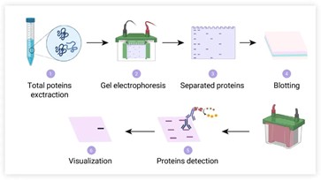
Which detection methods are compatible with membrane regeneration?
Compatible Detection Systems:
ECL Chemiluminescence System (Enhanced Chemiluminescence): This is the most compatible detection method with antibody stripping buffers. ECL relies on HRP-catalyzed luminol luminescence, resulting in clean background and excellent regeneration after antibody stripping.
Incompatible Detection Systems:
Chromogenic Methods: Western detection using chromogenic reagents such as DAB (3,3'-Diaminobenzidine), NBT/BCIP (Nitro Blue Tetrazolium/5-Bromo-4-Chloro-3-Indolyl Phosphate) is not suitable for membrane regeneration, as dyes form covalent bonds or precipitates with the membrane or proteins that cannot be eluted by stripping buffers.
How to maximize membrane regeneration efficiency and protect valuable samples?
Detection Order Strategy:
It is recommended to detect low-abundance target proteins first, then detect high-abundance proteins (e.g., housekeeping proteins) after stripping. Reasons include:
- Strong signals from high-abundance proteins (e.g., housekeeping) may leave "memory" on the membrane
- Detecting low-abundance proteins first avoids masking of weak signals by strong signals
- High expression of housekeeping proteins ensures successful detection even if regeneration is incomplete
Reuse Cycles:
Although membranes can theoretically be reused 2–3 times, each regeneration causes slight protein damage. Recommendations:
- First use: Detect the most critical target protein
- Second use: Detect housekeeping proteins for normalization
- Third use: Condition optimization or validation experiments
Protein Loss Monitoring:
After multiple regenerations, partial protein loss from the membrane may reduce signal intensity. Ponceau S staining can be used to check the integrity of protein bands before and after each regeneration.
Key Precautions for Use
Safety Protection:
- Mild and strong alkaline stripping buffers are slightly corrosive; always wear lab coats and disposable gloves during operation
- Acidic stripping buffer (pH=2.0) is also corrosive; avoid contact with skin and eyes
- All operations should be performed in a well-ventilated laboratory environment
Stripping Buffer Residue:
- Incompletely washed stripping buffer severely interferes with subsequent antibody binding, causing weak signals or high background
- Increase washing frequency or duration (especially after using strong alkaline stripping buffer)
- After washing, check membrane pH with pH paper to ensure it returns to neutral range
Blocking Buffer Selection:
Select the correct blocking buffer based on the enzyme conjugated to the secondary antibody. Incorrect blocking buffer (e.g., non-fat milk for AP system) causes high background or false-negative results.
Membrane Drying:
Avoid complete drying of the membrane during regeneration, as drying causes irreversible protein binding and membrane embrittlement. Keep the membrane moist when transferring between steps.
Sample Value:
- For extremely valuable samples (e.g., clinical specimens, primary cells), validate regeneration conditions with preliminary experiments first
- When using a new brand of stripping buffer, test regeneration efficiency with housekeeping antibodies to confirm protein retention and antibody stripping efficiency before formal experiments
By rationally selecting the type of primary and secondary antibody stripping buffer and strictly following standardized protocols, researchers can transform limited protein samples into multi-dimensional, reproducible Western Blot data, greatly improving experimental efficiency and ensuring accurate and comparable results.
Absin Recommended Primary and Secondary Antibody Stripping Buffers
| Cat. No. | Product Name | Size |
|---|---|---|
| abs969 | Western Blot Primary and Secondary Antibody Stripping Buffer (Neutral) | 500mL |
| abs968 | Western Blot Primary and Secondary Antibody Stripping Buffer (Mild Alkaline) | 500mL |
| abs967 | Western Blot Primary and Secondary Antibody Stripping Buffer (Strong Alkaline) | 500mL |
| abs958 | Western Blot Primary and Secondary Antibody Stripping Buffer (Acidic) | 500mL |
Contact Absin
Absin provides antibodies, proteins, ELISA kits, cell culture, detection kits, and other research reagents. If you have any product needs, please contact us.
| Absin Bioscience Inc. worldwide@absin.cn |
 Follow us on Facebook: Absin Bio Follow us on Facebook: Absin Bio |