- Cart 0
- English
How to realize the visualization and quantitative analysis of intracellular free cholesterol by using Filipin
April 24, 2026
Clicks:69
Filipin III is a polyene antibiotic that specifically binds to unesterified cholesterol. Upon excitation by ultraviolet light (excitation wavelength: 340-380 nm), the Filipin-cholesterol complex emits characteristic fluorescence (emission wavelength: 385-470 nm), enabling direct visualization and quantitative analysis of intracellular unesterified cholesterol. Compared with other cholesterol detection methods, Filipin staining features simple operation and high specificity, making it suitable for rapid detection of unesterified cholesterol in cultured cells and frozen tissue sections in vitro. It is a vital tool for investigating cholesterol homeostasis in cholesterol metabolism, lysosomal storage disorders, atherosclerosis, and neurodegenerative diseases.
Schematic diagram of Filipin III binding to cholesterol
Why does Filipin specifically recognize unesterified cholesterol?
Filipin belongs to the polyene macrolide compounds. The conjugated double bond system in its molecular structure can tightly bind to the sterol backbone of unesterified cholesterol through hydrophobic interactions and van der Waals forces, forming a stable complex. This binding is highly specific:
Selective Binding:
- Unesterified Cholesterol: Filipin only binds to unesterified cholesterol and does not bind to cholesterol esters (CE) or esterified cholesterol
- Steric Hindrance Sensitivity: Due to its large molecular size, Filipin cannot easily penetrate the tightly packed core of cholesterol esters, thus specifically labeling membrane structures or highly accessible unesterified cholesterol pools
Fluorescence Properties:
- Excitation Wavelength: 340-380 nm (UV range, commonly 360-380 nm)
- Emission Wavelength: 385-480 nm (blue fluorescence, peak at ~470-480 nm)
- Filter Set: DAPI or UV filter set can be used for observation
- Color Appearance: Blue or cyan fluorescence depending on microscope filter configuration
Association between cholesterol metabolism and diseases
What research fields can Filipin staining be applied to?
Lysosomal Storage Disorders and Cholesterol Transport Defects:
Filipin is a classic tool for diagnosing lysosomal storage disorders such as Niemann-Pick Type C (NPC). In NPC cells, unesterified cholesterol abnormally accumulates in lysosomes/endosomes due to cholesterol transport defects, and Filipin staining clearly reveals this "cholesterol accumulation" phenotype.
Neurodegenerative Disease Research:
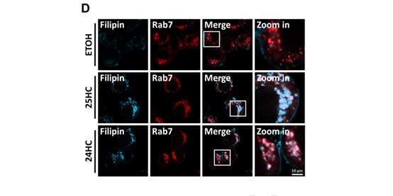
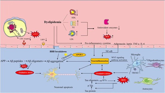
In Alzheimer's disease research, Filipin is used to reveal how APOE4-associated cholesterol metabolic abnormalities lead to microglial dysfunction and increased Aβ production. Changes in cholesterol homeostasis in astrocytes and neurons can also be intuitively visualized by Filipin staining.
Atherosclerosis and Foam Cell Formation:
Macrophages transform into foam cells after phagocytosing oxidized low-density lipoprotein, characterized by cholesterol accumulation. Filipin staining validates the pathological signaling axis of "macropinocytosis → cholesterol accumulation in foam cells → inflammasome activation".
Basic Research on Cellular Cholesterol Homeostasis:
Used to observe the effects of drug treatment (e.g., statins) or gene knockdown/overexpression (e.g., DHCR24, LXR, etc.) on intracellular cholesterol distribution and content.
How to establish a standardized staining protocol?
Preparation of Stock and Working Solutions
Stock Solution (1 mg/mL):
Dissolve solid powder in anhydrous DMSO to prepare a 1 mg/mL stock solution. Aliquot immediately into small volumes (e.g., 10-20 μL) and store at -20°C protected from light. Critical Tip: Stock solution is unstable; use within 1 month and avoid repeated freeze-thaw cycles.
Working Solution (0.1 mg/mL):
Dilute the stock solution to the desired working concentration (commonly 0.1 mg/mL) with PBS before use. Note: Working solution is extremely unstable; prepare fresh immediately before use, and all staining procedures must be performed strictly protected from light.
Cell Sample Staining Steps
Sample Preparation:
- Seed cells in an appropriate culture plate (e.g., 96-well plate, 3×10⁴ cells/well) and culture overnight
- Treat cells with compounds as per experimental design (e.g., 48-72 hours of drug treatment)
Staining Protocol:
- Washing: Discard the culture medium and gently wash cells with PBS 3 times
- Fixation: Add 4% paraformaldehyde and fix at room temperature for 20 minutes
- Washing: Wash cells with PBS 3 times after fixation
- Permeabilization (Optional): For observing cholesterol in organelles such as lysosomes and endoplasmic reticulum, treat with 0.1% Triton X-100 at room temperature for 5 minutes, then wash with PBS 3 times. Permeabilization is generally not required if only observing cholesterol on the cell membrane surface or in large lipid droplets
- Staining: Add an appropriate amount of Filipin III working solution (e.g., 0.1 mg/mL) and incubate at room temperature protected from light for 30-60 minutes (extend to 2 hours if cholesterol content is low)
- Washing: Discard the staining solution and gently wash cells with PBS 3 times to thoroughly remove unbound dye
- Observation: Perform fluorescence microscopy immediately
Tissue Section Staining (Fresh Frozen Sections)
Section Preparation:
Flash-freeze fresh tissue (e.g., mouse liver) with liquid nitrogen, embed in OCT compound, and cut 10 μm-thick sections using a cryostat, then mount on glass slides.
Staining Protocol:
- Equilibration and Washing: Equilibrate sections at room temperature, add PBS to cover the tissue, tilt to drain, repeat 3 times. Blot excess liquid from the edge with filter paper
- Staining: Add Filipin III working solution (e.g., 0.1 mg/mL) to cover the tissue and incubate at room temperature protected from light for 30-60 minutes
- Washing: Gently wash sections with PBS 3 times, 5 minutes each
- Mounting and Observation: Blot excess liquid, mount with anti-fade mounting medium, and examine immediately under a microscope
How to set up control experiments to verify staining specificity?
To ensure the specificity and reliability of experimental results, the following controls are recommended:
| Control Type | Procedure | Purpose |
|---|---|---|
| Negative Control | Treat cells with Methyl-β-cyclodextrin (5-10 mM, incubate at 37°C for 30-60 min) to extract cholesterol before staining | Verify that fluorescence signal originates from cholesterol |
| Positive Control | Treat cells with U-18666A (1.25 μM, incubate at 37°C for 48 h) to block cholesterol transport and induce lysosomal cholesterol accumulation | Validate the effectiveness of the staining system |
| Dye Control | Incubate samples with PBS/buffer only without Filipin III | Exclude autofluorescence interference from samples or buffers |
Key Precautions and Troubleshooting
Strict Light Protection:
Filipin III is extremely light-sensitive; all operations (dissolution, staining, storage) must be performed protected from light. Its fluorescence is highly prone to photobleaching; use the lowest excitation light intensity and shortest exposure time during imaging, and prioritize high-sensitivity cameras.
Solution Stability:
Both stock and working solutions are unstable; aliquot for storage, avoid repeated freeze-thaw cycles, and use as soon as possible. Discard the stock solution if it becomes turbid or yellowish, indicating degradation.
Concentration and Incubation Time Optimization:
Optimal staining concentration and time vary for different cells or tissues. Preliminary optimization is recommended:
- No Fluorescence Signal: May result from degraded Filipin III (check if powder is yellow) or low cholesterol content; extend staining time (to 2 hours) or increase working solution concentration appropriately
- High Background Fluorescence: Insufficient washing or non-specific binding; increase washing frequency and duration, or use PBS containing 1-5% BSA as diluent/wash buffer
- Weak Signal or Rapid Photobleaching: Reduce excitation light intensity immediately and use high-speed cameras to shorten single exposure time
Microscope Parameter Settings:
- Objective Recommendation: 40× or 63× oil immersion objective
- Excitation Wavelength: 340-380 nm (UV range, commonly 360-380 nm)
- Emission Wavelength: 385-480 nm (blue fluorescence, commonly 420-470 nm or DAPI channel)
- Filter Set: DAPI or UV filter set
Only Plasma Membrane Signal with Weak Intracellular Signal:
May be due to high plasma membrane cholesterol background; try brief pretreatment with Methyl-β-cyclodextrin (optimize time) to partially extract plasma membrane cholesterol and expose intracellular cholesterol pools.
By strictly following light-protected operations, optimizing staining conditions, and setting appropriate controls, Filipin staining provides reliable and intuitive visualization data for cholesterol metabolism research, helping to elucidate the pathological mechanisms of cholesterol homeostasis dysregulation in various diseases.
Absin Filipin Recommendation
| Cat. No. | Product Name | Size |
|---|---|---|
| abs42018484 | Filipin III | 500ug/1mg |
Contact Absin
Absin provides antibodies, proteins, ELISA kits, cell culture, detection kits, and other research reagents. If you have any product needs, please contact us.
| Absin Bioscience Inc. worldwide@absin.cn |
 Follow us on Facebook: Absin Bio Follow us on Facebook: Absin Bio |