- Cart 0
- English
How to accurately select nucleic acid dyes according to experimental scenarios?
April 22, 2026
Clicks:78
In molecular biology and cell biology experiments, nucleic acid dyes are indispensable tools for visualizing DNA and RNA. From classical ethidium bromide (EB) to next-generation safety dyes, and further to fluorescent probes specifically designed for cell viability detection, different types of nucleic acid dyes exhibit fundamental differences in their mechanisms of action, spectral properties, and application scenarios. How to make the correct choice between gel electrophoresis and cell staining? What are the technical advantages and disadvantages of red versus green dyes? This article systematically analyzes the technical characteristics and experimental adaptation strategies of three mainstream categories of nucleic acid dyes.
What Are the Fundamental Differences in Their Mechanisms of Action?
Based on chemical properties and modes of action, commonly used nucleic acid dyes in the laboratory can be classified into two major categories:
Category I: Gel Electrophoresis Dyes
These dyes intercalate between the base pairs of nucleic acid duplexes, producing fluorescent signals upon excitation at specific wavelengths. They are suitable for visualization of nucleic acid bands following agarose gel or polyacrylamide gel electrophoresis, and are capable of detecting double-stranded DNA (dsDNA), single-stranded DNA (ssDNA), and RNA. These dyes possess lipophilic macromolecular structural characteristics and cannot penetrate intact cell membranes; therefore, they are primarily used for detection of ex vivo nucleic acid samples.
Category II: Cell Staining Dyes
Unlike gel dyes, these dyes are specifically designed for labeling intracellular nucleic acids. They readily traverse damaged cell membranes but cannot permeate intact plasma membranes of viable cells, making them ideal probes for distinguishing live cells from dead cells. Upon binding to nucleic acids, fluorescence intensity can increase by more than 500-fold, making them suitable for multicolor fluorescence analysis platforms such as fluorescence microscopy and flow cytometry.
How to Choose Between Red and Green Dyes in Nucleic Acid Gel Electrophoresis?
In the field of nucleic acid gel electrophoresis, red and green dyes represent two distinct technical approaches, both aiming to replace highly toxic and strongly mutagenic ethidium bromide (EB), yet differing in spectral properties and instrument compatibility:
Red-Series Dyes (EB-Spectrum Analogs)
- Spectral Properties: Excitation wavelength approximately 300 nm (UV light), emitting red fluorescence
- Instrument Compatibility: Directly observable using standard EB filter sets or SYBR filter sets without modification of existing UV gel transilluminators
- Sensitivity: Minimal impact on nucleic acid migration, suitable for electrophoretic staining of various fragment sizes
- Stability: Can be directly added during microwave heating of agarose gel preparation; extremely stable in acidic or alkaline buffers; high photostability
- Limitations: Cannot be fully excited by 488 nm argon-ion lasers or similar wavelength visible light sources; not recommended for such imaging systems
Green-Series Dyes (SYBR Green I-Spectrum Analogs)
- Spectral Properties: Excitation wavelength compatible with blue light or UV light, emitting green fluorescence
- Instrument Compatibility: Same spectral observation position as EB; compatible with standard EB filter sets or SYBR filter sets; also compatible with blue light imaging systems, allowing observation under visible light
- Sensitivity: Fluorescence brightness exceeds that of EB by more than tenfold, with visibly brighter bands observable to the naked eye
- Stability: Stable for 6 months under routine laboratory lighting conditions; extremely stable in acidic or alkaline buffers at room temperature
- Advantages: Suitable for laser imaging systems; optimal choice for experimental scenarios requiring blue light excitation
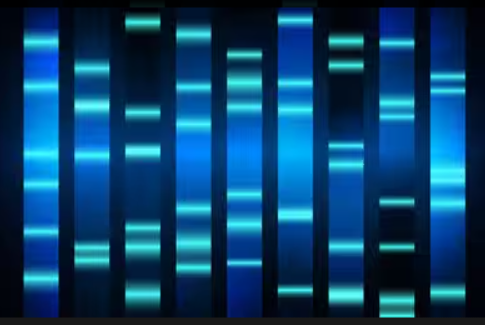
Gel Electrophoresis Results Display
Why Must Dedicated Nucleic Acid Dyes Be Used in Cell Experiments?
In cell viability assays, cytotoxicity evaluation, or flow cytometry experiments, dedicated nucleic acid dyes capable of distinguishing live from dead cells must be employed. The core characteristic of these dyes is membrane permeability selectivity:
Mechanism of Action
Dye molecules recognize dead cells with compromised membrane integrity, enter the nucleus to bind DNA, and produce intense fluorescence; whereas intact plasma membranes of viable cells prevent dye entry, maintaining a non-fluorescent state.
Spectral Advantages
Typical green dead cell dyes exhibit spectral characteristics of 502 nm excitation/525 nm emission, fully compatible with FITC filter sets, and are applicable to standard fluorescence microscopes, fluorescence microplate readers, and flow cytometers.
Multicolor Compatibility
Due to emission in the green spectral band, these dyes can be combined with other fluorescent probes in the red and blue channels (such as PI, DAPI) to achieve multiparametric cellular analysis.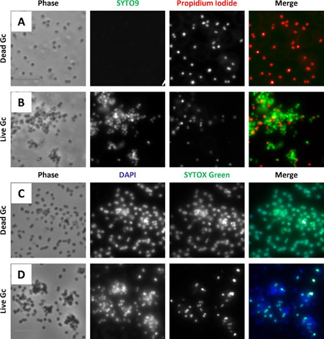
SYTOX Green Dead Cell Staining Effect
Standard Operating Procedures for Three Experimental Scenarios
Scenario I: Pre-Electrophoresis Staining of Agarose Gels (GelCast Method)
This is the recommended standard method, featuring simple operation and minimal dye consumption:
- Add nucleic acid dye directly during gel preparation (typically diluted 2,000-fold from 10,000× stock solution)
- Dye can be added directly to hot agarose solution without cooling; mix thoroughly and cast the gel
- Perform electrophoresis according to standard protocols; dye does not degrade during electrophoresis
- After electrophoresis, visualize directly under UV or blue light transilluminator without destaining or rinsing
Scenario II: Post-Electrophoresis Staining of Agarose Gels (Soak Method)
Suitable for precast polyacrylamide gels or situations requiring optimized staining results:
- Complete electrophoresis according to standard protocols
- Dilute dye approximately 3,300-fold in 0.1 M NaCl solution to prepare 3× staining solution
- Carefully immerse the gel in staining solution and incubate with agitation at room temperature for approximately 30 minutes (adjust according to gel thickness and agarose concentration; polyacrylamide gels typically require 30 minutes to 1 hour)
- Staining solution can be recovered and reused 2-3 times
Scenario III: Live/Dead Cell Staining
For flow cytometry or fluorescence microscopy observation:
- Adherent cells can be stained directly on slides; suspension cells require centrifugation, supernatant removal, and resuspension in buffer
- Staining concentration should be optimized according to cell type: typically 0.5-5 μM for bacteria, 1-50 μM for yeast, and 10 nM-1 μM for eukaryotic cells
- Incubation time: minimum 5 minutes for bacteria, minimum 10 minutes for yeast and eukaryotic cells
- After staining, dead cells with bright green fluorescence can be directly observed under fluorescence microscope, or quantitatively analyzed using flow cytometry
Safety and Operational Considerations
Safety Advantages
Next-generation gel dyes have been validated by the Ames test as non-mutagenic. Their lipophilic macromolecular characteristics prevent them from penetrating cell membranes, offering significantly higher operational safety compared to EB.
Key Operational Points:
- Avoid Repeated Freeze-Thaw Cycles: Dye solutions should be aliquoted and stored to prevent activity loss
- Protect from Light: Fluorescent dyes are inevitably subject to photobleaching; protect from light during preparation and use
- Use Plastic Ware: Use plastic tubes for diluting staining solutions to avoid concentration deviations caused by glass adsorption
- Avoid Phosphate Buffers: Certain cell staining applications do not recommend use of phosphate-containing buffers; ensure tubes are thoroughly cleaned to avoid background interference
- Personal Protection: Although toxicity is low, wearing a lab coat and disposable gloves is still recommended during operation
Staining Optimization Recommendations:
- If bands appear diffuse or separation is suboptimal, try reducing agarose concentration, using a longer gel, or extending electrophoresis time
- For smearing of restriction enzyme-digested DNA samples, try alternative staining methods or adjust electrophoresis conditions
- If resolution decreases after plasmid extraction, check whether RNase treatment is adequate or try the soak staining method
Conclusion
From gel electrophoresis to cell viability detection, the choice of nucleic acid dye directly impacts the accuracy of experimental data and operator safety. Red-series dyes are suitable for laboratories continuing to use traditional UV imaging equipment, green-series dyes provide better compatibility for blue light imaging and laser scanning, while dedicated dead cell dyes are indispensable in cell biology research. Understanding the spectral properties, membrane permeability differences, and optimal operating conditions of various dyes is a prerequisite for obtaining clear and reliable nucleic acid signals. As fluorescence imaging technology continues to advance, next-generation safe, sensitive, and multifunctional nucleic acid dyes will continue to drive life science research toward higher resolution and higher throughput.
Absin Nucleic Acid Dye Recommendations
| Cat. No. | Product Name | Specification |
|---|---|---|
| abs9181 | GelRed Nucleic Acid Gel Stain | 1mL/1mL×5 |
| abs9921 | GelGreen Nucleic Acid Gel Stain | 500μL |
| abs47038985 | SYTOX Green Dead Cell Nucleic Acid Stain | 500μL |
Contact Absin
Absin provides antibodies, proteins, ELISA kits, cell culture, detection kits, and other research reagents. If you have any product needs, please contact us.
| Absin Bioscience Inc. worldwide@absin.cn |
 Follow us on Facebook: Absin Bio Follow us on Facebook: Absin Bio |