- Cart 0
- English
Precious and hard-to-measure samples, missing spatial information! Is research on human early embryonic gastrulation facing bottlenecks? Absin Multicolor Kit Helps Unlock Developmental Secrets in *Nature Cell Biology*
April 17, 2026
Clicks:69
In human embryonic development research, gastrulation represents a critical juncture for establishing the body axes and initiating trilaminar germ layer differentiation. However, studies in this field have been long constrained by the scarcity of early embryonic specimens and the lack of spatial dimension information. Recently, a study published in Nature Cell Biology achieved the first comprehensive characterization of the spatial cellular atlas and molecular mechanisms of a Carnegie stage (CS) 7 human embryo through the innovative integration of spatial transcriptomics and multiplex immunofluorescence validation—and Absin's multicolor reagent kits served as the critical support for the immunofluorescence validation component of this research.
Title:Spatial transcriptomic characterization of a Carnegie stage 7 human embryo
Journal:Nature Cell Biology (IF 19.1)
DOI:https://doi.org/10.1038/s41556-024-01597-3
Key Reagents:Seven-Color Multiplex Fluorescence Immunohistochemistry Staining Kit(abs50015), Antibody Stripping Buffer (abs994)
I. Research Strategy: Spatial Dimension + Molecular Validation to Overcome Early Embryo Research Bottlenecks
While conventional single-cell transcriptomics can resolve cell types, it fails to reconstruct the spatial positioning of cells within the embryo, leading to challenges in distinguishing mesodermal subtypes and identifying critical signaling centers. This study innovatively employed a technical pipeline combining "spatial transcriptomics (Stereo-seq) + multiplex immunofluorescence validation":
- Eighty-two serial cryosections were prepared from a CS7 human embryo, and Stereo-seq technology was utilized to acquire spatial transcriptomic data at single-cell resolution;
- Transcriptomic and spatial information were integrated for clustering analysis and 3D embryonic model reconstruction;
- Multiplex immunofluorescence experiments were performed to validate key cell types (e.g., AVE, PGCs) and molecular marker expression localization, with core staining reagents sourced from Absin products;
- Regulatory network analysis and pseudotime analysis were combined to elucidate cell specification and signaling communication mechanisms during gastrulation.
II. Core Research Findings: Advancing Our Understanding of Human Early Embryonic Development
1. 3D Reconstruction of CS7 Embryo Spatial Atlas Defining 11 Major Tissue Clusters
Through section sequencing and data integration, the study successfully constructed a 3D model of the CS7 embryo, precisely identifying 11 major tissue clusters including ectoderm, mesoderm, endoderm, amnion, and yolk sac, clearly presenting the distribution patterns of cells along the anteroposterior and dorsoventral axes (Original Figures 1a, 1b). This achievement resolved the limitation of transcriptome-only approaches in distinguishing spatially specific cell subtypes.
2. Confirmation of Early Mesodermal Subtype Specification with Defined Spatial Localization Differences
The study revealed that distinct mesodermal subtypes, including paraxial mesoderm and lateral plate mesoderm, already exist in the CS7 embryo. Although these subtypes share classical markers (e.g., TBXT, MESP1), their unique spatial positioning can be clearly defined through spatial information (Original Figures 2d, 2e), correcting the traditional perception that "mesoderm remains unspecialized at this stage."
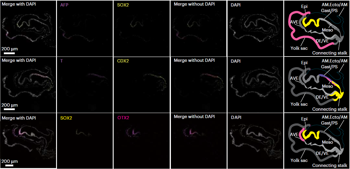
3. First Confirmation of AVE Existence in Human Embryos Revealing Axis Establishment Mechanisms
Through immunofluorescence staining validation (Original Figure 3f), the study definitively established the existence and anterior-central localization of the anterior visceral endoderm (AVE) in human CS7 embryos. The AVE regulates anteroposterior axis establishment through secretion of signaling molecules including BMP and FGF, with markers OTX2 and DKK1 clearly detectable following staining with Absin reagents, providing critical evidence for the conservation of axis formation mechanisms across species.
4. Localization of Primordial Germ Cells Tracing Developmental Origins
The study identified primordial germ cells (PGCs) in the connecting stalk region, confirmed through SOX17 and TFAP2C double-positive staining (Original Figure 4d), and hypothesized that PGCs may originate from ectoderm-derived cells, providing spatial reference for germ cell development research.
5. Discovery of Yolk Sac Hematopoiesis Independent of Hematopoietic Stem Cells
In the yolk sac region, the study identified 10 hematopoiesis-related subpopulations including hemogenic endothelial progenitors and erythroid precursors, confirming the initiation of hematopoietic stem cell-independent hematopoiesis at the CS7 stage (Original Figures 5d, 5e), offering novel perspectives for investigating the origins of hematopoietic development.
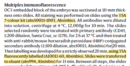
III. Absin Products: The "Reliable Partner" for Immunofluorescence Validation
The core validation component of this study—multiplex immunofluorescence experiments—critically depended on two Absin core products to ensure staining signal specificity and stability:
1. Product List and Application Scenarios
| Catalog No. | Product Name | Application in This Study |
|---|---|---|
| abs50015 | Seven-Color Multiplex Fluorescence Immunohistochemistry Staining Kit | Used for multiplex marker staining on tissue sections, enabling simultaneous detection of multiple targets |
| abs994 | Antibody Stripping Buffer |
2. Core Functions of Products in This Study
3. Key Image-Text Correspondence
 Original Figure 3f: Immunofluorescence staining results for OTX2 protein, confirming AVE localization in the anterior-central region of the embryo. Staining was performed using Absin HRP secondary antibody (abs50015) and TSA kit (abs50015-100T), with clear signals and precise localization.
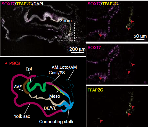 Original Figure 4d: SOX17 and TFAP2C double-positive staining identifying PGCs, enabled by the multiplex staining capability of the TSA kit for simultaneous detection of both markers.
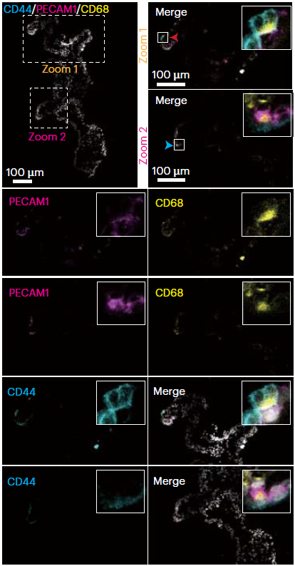 Original Figure 5e: Co-staining of PECAM1, CD68, and CD44 in yolk sac tissue validating hematopoiesis-related cell localization; Absin products ensured signal discrimination and stability for multi-target staining.
IV. Conclusion: Absin Empowering Scientific Breakthroughs and Life Science Exploration
Every advance in human early embryonic development research depends on precise and reliable experimental tools. Through the innovative combination of spatial transcriptomics and immunofluorescence, this study has transformed our understanding of gastrulation, with Absin's TSA multiplex staining kits, antibody stripping buffer (abs994), and HRP secondary antibodies serving as the "unsung heroes" behind this breakthrough—from signal amplification to multiplex compatibility, from sample conservation to result stability, comprehensively meeting the demanding requirements of precious specimen experiments.
Moving forward, Absin will continue to deepen its commitment to life science reagents, supporting more frontier research with superior products, empowering scientists to unravel the core mysteries of life development, and advancing progress in human health and regenerative medicine!
Products Used in This Study:
| Catalog No. | Product Name | Size |
|---|---|---|
| abs50015 | Seven-Color Multiplex Fluorescence Immunohistochemistry Staining Kit (Mouse/Rabbit Universal Secondary Antibody) | 20T/50T/100T |
| abs994 | Antibody Stripping Buffer (mIHC专用) | 30 mL |
More Multiplex Fluorescence Immunohistochemistry Kits
| Catalog No. | Product Name | Size |
|---|---|---|
| abs50086 | Two-Color Multiplex Fluorescence Immunohistochemistry Staining Kit (Anti-Rabbit Secondary Antibody) | 100T |
| abs50087 | Two-Color Multiplex Fluorescence Immunohistochemistry Staining Kit (Mouse/Rabbit Universal Secondary Antibody) | 100T |
| abs50088 | Three-Color Multiplex Fluorescence Immunohistochemistry Staining Kit (Anti-Rabbit Secondary Antibody) | 100T |
| abs50089 | Three-Color Multiplex Fluorescence Immunohistochemistry Staining Kit (Mouse/Rabbit Universal Secondary Antibody) | 100T |
| abs50012 | Four-Color Multiplex Fluorescence Immunohistochemistry Staining Kit (Mouse/Rabbit Universal Secondary Antibody) | 20T/50T/100T |
| abs50168 | Four-Color Multiplex Fluorescence Immunohistochemistry Staining Kit B (Anti-Rabbit Secondary Antibody) | 20T/50T/100T |
| abs50013 | Five-Color Multiplex Fluorescence Immunohistochemistry Staining Kit (Mouse/Rabbit Universal Secondary Antibody) | 20T/50T/100T |
| abs50029 | Five-Color Multiplex Fluorescence Immunohistochemistry Staining Kit (Anti-Rabbit Secondary Antibody) | 20T/50T/100T |
| abs50014 | Six-Color Multiplex Fluorescence Immunohistochemistry Staining Kit (Mouse/Rabbit Universal Secondary Antibody) | 20T/50T/100T |
| abs50030 | Six-Color Multiplex Fluorescence Immunohistochemistry Staining Kit (Anti-Rabbit Secondary Antibody) | 20T/50T/100T |
| abs50048 | Six-Color Multiplex Fluorescence Immunohistochemistry Staining Kit (Plus) (Anti-Rabbit Secondary Antibody) | 20T/50T/100T |
| abs50049 | Six-Color Multiplex Fluorescence Immunohistochemistry Staining Kit (Plus) (Mouse/Rabbit Universal Secondary Antibody) | 20T/50T/100T |
| abs50015 | Seven-Color Multiplex Fluorescence Immunohistochemistry Staining Kit (Mouse/Rabbit Universal Secondary Antibody) | 20T/50T/100T |
| abs50031 | Seven-Color Multiplex Fluorescence Immunohistochemistry Staining Kit (Anti-Rabbit Secondary Antibody) | 20T/50T/100T |
| abs50037 | Seven-Color Multiplex Fluorescence Immunohistochemistry Staining Kit (Plus) (Mouse/Rabbit Universal Secondary Antibody) | 20T/50T/100T |
| abs50038 | Seven-Color Multiplex Fluorescence Immunohistochemistry Staining Kit (Plus) (Anti-Rabbit Secondary Antibody) | 20T/50T/100T |
| abs50165 | Seven-Color Multiplex Fluorescence Immunohistochemistry Staining Kit (770 Dye Enhanced Version) (Anti-Rabbit Secondary Antibody) | 20T/50T/100T |
| abs50166 | Seven-Color Multiplex Fluorescence Immunohistochemistry Staining Kit (770 Dye Enhanced Version) (Mouse/Rabbit Universal Secondary Antibody) | 20T/50T/100T |
| abs50018 | Ten-Color Multiplex Fluorescence Immunohistochemistry Staining Kit | 100T |
| abs50083 | Lung Cancer Tumor Microenvironment Multiplex Fluorescence Immunohistochemistry Detection Kit (I) | 20T |
| abs50084 | Lung Cancer Tumor Microenvironment Multiplex Fluorescence Immunohistochemistry Detection Kit (II) | 20T |
Limited-Time Offer! Absin Multicolor Services Starting at 200 RMB per Four-Color Section (50% Discount)
Click for Details[Disclaimer]This article is based on the original publication in Nature Cell Biology (DOI: 10.1038/s41556-024-01597-3) and has been compiled and interpreted by AI. All intellectual property rights to the original figures, data, and content belong to the original journal and the research team. Should any infringement occur, please contact us promptly for removal, and we will cooperate fully to resolve the matter.
Contact Absin
Absin provides antibodies, proteins, ELISA kits, cell culture, detection kits, and other research reagents. If you have any product needs, please contact us.
| Absin Bioscience Inc. worldwide@absin.cn |
 Follow us on Facebook: Absin Bio Follow us on Facebook: Absin Bio |