- Cart 0
- English
Why Can't You Get Intact Structures from Your Bone Tissue Sections?
March 18, 2026
Clicks:73
In histopathology laboratories, bone tissue sectioning is widely regarded as one of the most technically challenging procedures. When you place fixed bone tissue blocks into the paraffin embedding station, anticipating perfect sections for morphological observation, you often encounter frustration during the sectioning phase: microtome blades either develop nicks from the hard calcified matrix, or the tissue shatters beneath the cutting edge, ultimately yielding sections with uneven thickness and severe fragmentation. The root cause of this dilemma lies in a critical yet frequently overlooked step—decalcification. And EDTA decalcifying solution is precisely the "gentle yet powerful tool" that resolves this challenge.
What Exactly Is EDTA Decalcifying Solution?
EDTA decalcifying solution is a chelating-type decalcification reagent based on ethylenediaminetetraacetic acid (EDTA), specifically designed for removing inorganic calcium salts from bone tissue and calcified lesions to enable subsequent paraffin embedding and routine histological sectioning. Unlike conventional acidic decalcifying agents, EDTA forms stable chelates with calcium ions, "dissolving" calcium in a gentle manner rather than through the erosive destruction of bone matrix by strong acids.
Standard EDTA decalcifying solution typically comprises the following components:
- EDTA: The core decalcifying component, which forms hexadentate chelation rings with Ca²⁺ through its four carboxyl groups and two amino groups. This binding exhibits an extremely high stability constant (log Kf ≈ 10.7), effectively "capturing" both free calcium in tissue and calcium ions within hydroxyapatite crystals.
- Buffer System: Maintains pH at 7.2-7.4 in the neutral to weakly alkaline range. This pH environment ensures EDTA chelating activity while maximizing preservation of tissue antigenicity and enzymatic activity.
- Some formulations contain formalin: Enables simultaneous fixation and decalcification, reducing tissue damage.
Depending on application scenarios, EDTA decalcifying solutions are classified as standard EDTA type and formalin-EDTA composite type. The former offers relatively faster decalcification speed, while the latter provides superior tissue structural preservation at the cost of slower processing.
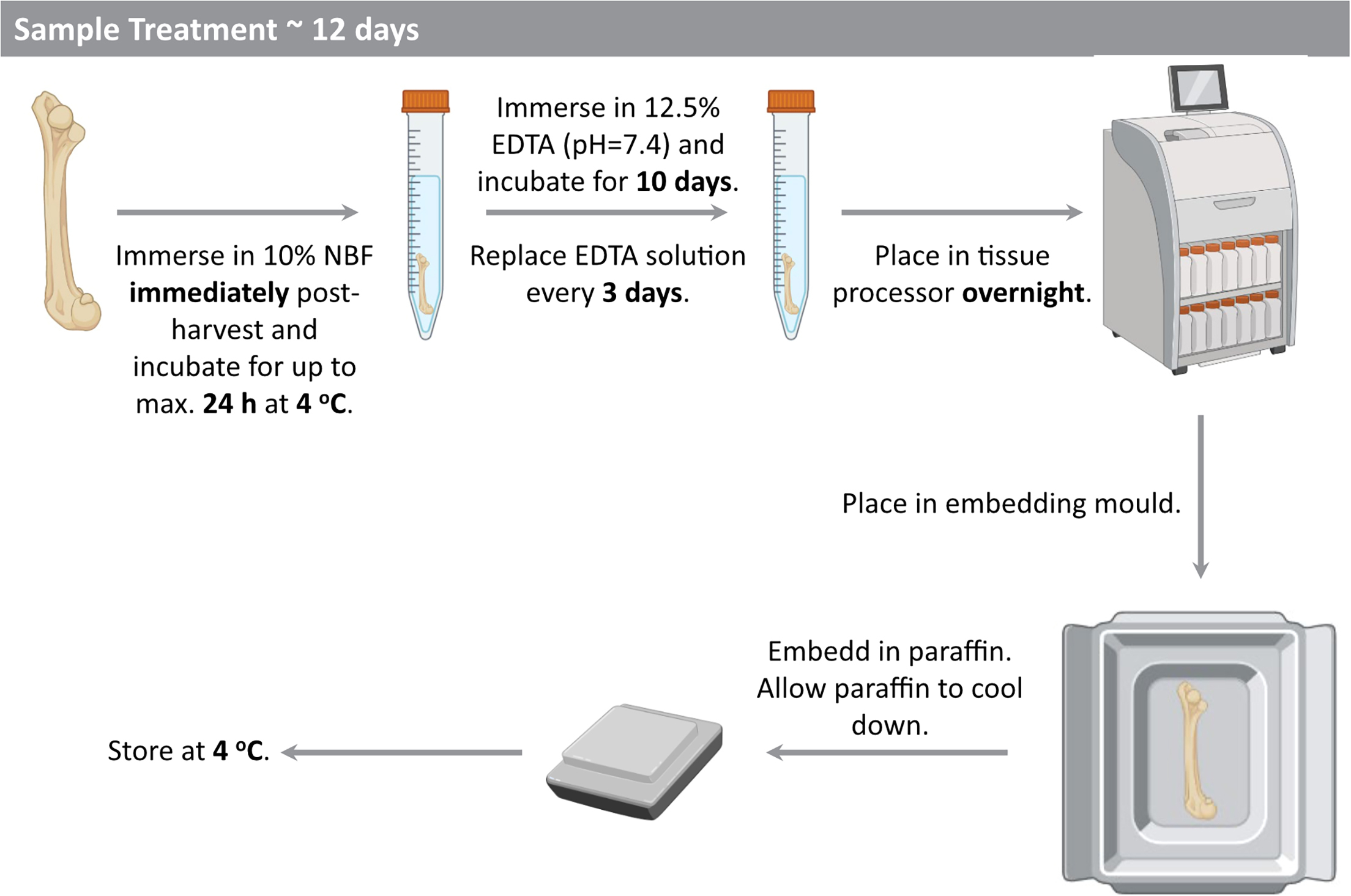
Figure 1: Standard workflow from bone tissue sampling to sectioning. After fixation, tissue must be immersed in EDTA decalcifying solution for several weeks to months until calcium is completely removed, followed by dehydration, clearing, paraffin infiltration, and final embedding and sectioning. The thoroughness of decalcification directly determines section quality and subsequent staining results.
Why Is EDTA Known as the "Gentlest Decalcifying Agent"?
Among decalcification technologies, EDTA is not the fastest performer, yet its unique gentle characteristics have established it as the "gold standard" for bone tissue processing.
Chelation Mechanism: Selective Calcium Removal
EDTA decalcification operates through chelation chemistry rather than acid-base neutralization. It does not react with organic components in bone matrix (primarily collagen), but selectively targets calcium ions within hydroxyapatite crystals [Ca₁₀(PO₄)₆(OH)₂]. This selectivity means the organic scaffold of bone tissue remains intact, with extracellular matrix ultrastructure preserved. In contrast, acidic decalcifying agents such as nitric acid or formic acid simultaneously hydrolyze collagen, rendering tissue fragile and prone to fragmentation.
Neutral pH: Protection of Biological Macromolecules
Acidic decalcification environments (pH<2) cause protein denaturation, nucleic acid degradation, and loss of enzymatic activity. EDTA decalcifying solution maintains pH 7.2-7.4, approaching physiological conditions, which enables:
- Antigenic epitope preservation: Antigenic determinants required for immunohistochemical (IHC) staining remain intact
- Nucleic acid integrity: DNA and RNA degradation is minimized, supporting molecular pathology techniques such as in situ hybridization (ISH)
- Enzymatic activity preservation: Partial hydrolase activity is maintained for histochemical enzyme staining
Controllability: Precise Determination of Decalcification Endpoint
Although EDTA decalcification proceeds slowly (typically requiring several weeks to months), this "slowness" precisely provides controllability. Researchers can monitor decalcification progress in real-time through physical detection methods (needle penetration, manual palpation), avoiding the dilemma of both insufficient decalcification (sectioning difficulties) and over-decalcification (tissue damage).
Which Experimental Scenarios Absolutely Require EDTA Decalcifying Solution?
Bone Tissue Pathomorphological Studies
This represents the most classical application scenario. Whether diagnosing bone tumors (osteosarcoma, chondrosarcoma, bone metastasis) or evaluating metabolic bone diseases (osteoporosis, osteomalacia, hyperparathyroid bone disease), high-quality bone tissue sections are essential. Sections following EDTA decalcification clearly display trabecular bone structure, osteocyte morphology, marrow cavity components, and mineralization fronts, providing reliable evidence for pathological diagnosis.
Immunohistochemistry (IHC) and Immunofluorescence (IF)
When research objectives involve specific protein expression in bone tissue (such as osteocalcin, type I collagen, osteoclast marker TRAP, osteoblast transcription factor Osterix), or require labeling tumor cell origin markers, EDTA decalcification is virtually the only viable option. Acidic decalcification destroys most antigenic epitopes, leading to false-negative results; whereas EDTA-decalcified tissue sections exhibit high antigen retention, low background staining, and strong signal specificity.
In Situ Hybridization (ISH) and Fluorescence In Situ Hybridization (FISH)
These techniques rely on the integrity of nucleic acid sequences within tissue. Whether detecting viral DNA (such as CMV infection post-bone marrow transplantation), gene rearrangements (such as EWSR1-FLI1 fusion gene in Ewing sarcoma), or chromosomal abnormalities (such as myelodysplastic syndromes), EDTA decalcification maximally protects DNA and RNA from degradation, ensuring specific probe binding.
Bone Development and Regeneration Research
In bone biology research, observation of bone formation and bone resorption processes is frequently required. EDTA-decalcified bone tissue sections enable:
- Histochemical staining: Such as Von Kossa staining (displaying mineralization nodules), TRAP staining (displaying osteoclast activity)
- Immunohistochemical double staining: Simultaneous labeling of osteoblast and osteoclast markers to study cell-cell interactions
- Laser capture microdissection: Precise isolation of specific cell populations from complex bone tissue for transcriptomic analysis
Pathological Examination of Calcified Lesions
Beyond bone tissue, EDTA decalcifying solution is also applicable to calcified lesions in other anatomical locations:
- Atherosclerotic plaques: Calcium salt deposits within plaques damage microtome blades; EDTA decalcification yields intact vascular intima sections
- Calcific tendinitis: Hydroxyapatite deposits within tendons require decalcification processing
- Breast calcifications: Localization and characterization of microcalcifications
- Thyroid calcification: Observation of psammoma bodies
Spatial Transcriptomics and Multi-Omics Research
Emerging spatial omics technologies (such as Visium, Stereo-seq) require tissue sections with high structural integrity and RNA quality sufficient for in situ sequencing. The gentle characteristics of EDTA decalcification make it the preferred method for bone tissue spatial transcriptomics sample preparation. In contrast, acidic decalcification severely degrades RNA, limiting the application of these cutting-edge technologies.
How to Master the "Art" of EDTA Decalcification?
Control of Sampling Thickness
The optimal thickness for bone tissue blocks is approximately 5mm. Excessive thickness (>10mm) significantly prolongs decalcification time and may result in incomplete central decalcification; insufficient thickness (<3mm) shortens processing time but may cause tissue structure loosening due to over-decalcification. For irregular specimens such as long bones, longitudinal splitting or transverse sectioning to appropriate thickness is recommended.
Sequential Strategy for Fixation and Decalcification
Two operational pathways are available:
- Fixation followed by decalcification: The standard protocol for routine pathology—10% neutral formalin fixation for 48 hours followed by transfer to EDTA decalcification. Advantages include adequate fixation and stable tissue structure.
- Simultaneous fixation and decalcification: Using formalin-EDTA composite decalcifying solution, suitable for time-critical cases. Note that formalin fixation efficacy diminishes at alkaline pH, potentially requiring extended processing time.
Absolute prohibition: Decalcification before fixation will cause tissue autolysis within the decalcifying solution, resulting in severe cellular structural damage.
Precise Determination of Decalcification Endpoint
Physical detection methods provide practical means for assessing decalcification endpoints:
- Needle penetration test: Insert a syringe needle vertically into the thickest portion of tissue; if resistance is uniform without "scratchy" calcification sensation, decalcification is complete
- Manual palpation: Gently compress tissue edges with forceps or fingers; if tissue exhibits some elasticity rather than rock-hard consistency, decalcification may be terminated
- X-ray detection: The most accurate method, though requiring specialized equipment
Warning: Over-decalcification causes excessive tissue softening, section wrinkling, and uneven staining. Once over-decalcified, the process is irreversible.
Balance Between Temperature and Time
EDTA decalcification can proceed at room temperature or 37°C. Moderate temperature elevation (37-40°C) accelerates chelation reactions, shortening decalcification time by approximately 30%-50%, but vigilance is required:
- Temperature ceiling: Must not exceed 60°C, otherwise tissue protein denaturation and structural disintegration occur
- Microwave assistance: Special formulations support 200W microwave short heating (5 minutes per cycle, 3-5 minute intervals, 3-5 repetitions), significantly shortening decalcification time to several days, but strict control of power and duration is essential to prevent localized overheating
Importance of Solution Change Frequency
As EDTA binds calcium ions, it gradually becomes saturated and decalcification efficiency declines. Fresh decalcifying solution should be replaced weekly, or immediately when the solution becomes turbid or precipitates appear. Continuous stirring with a magnetic stirrer increases ion exchange efficiency and shortens decalcification time.
From "Slow and Steady" to "Meticulous Craftsmanship": The Technical Philosophy of EDTA Decalcification
In an era where "rapid results" has become the norm, the "slowness" of EDTA decalcification may seem incongruous. However, behind this slowness lies the ultimate pursuit of sample quality.
Acidic decalcification (such as nitric acid decalcification) can indeed complete processing within hours to days, but at the cost of:
- Nuclear pyknosis, enhanced cytoplasmic eosinophilia, and loss of morphological detail
- Significantly decreased immunohistochemical positivity rates, even false negatives
- Severe nucleic acid degradation, failure of molecular pathology detection
The weeks of waiting with EDTA decalcification yield:
- Clear cellular morphology with distinct nuclear-cytoplasmic contrast
- Sensitive and specific immunohistochemical staining with accurate localization
- Excellent nucleic acid integrity, supporting downstream PCR, sequencing, and other analyses
For challenging cases requiring precise pathological diagnosis, or experiments pursuing high-quality research data, the time investment of EDTA decalcification is entirely worthwhile.
Conclusion
EDTA decalcifying solution embodies the wisdom of "winning through slowness" in histopathological technology. In the processing of bone tissue and calcified lesions, it is not the fastest option, but the most reliable, gentle, and fidelity-preserving solution. From routine pathological diagnosis to cutting-edge spatial omics, from morphology to molecular pathology, EDTA decalcification technology remains the key to unlocking the microscopic world of hard tissues. Mastering the essence of this technology means achieving the optimal balance between hardness and softness, speed and quality, destruction and preservation.
Absin EDTA Decalcifying Solution Recommendations
Contact Absin
Absin provides antibodies, proteins, ELISA kits, cell culture, detection kits, and other research reagents. If you have any product needs, please contact us.
| Absin Bioscience Inc. worldwide@absin.cn |
 Follow us on Facebook: Absin Bio Follow us on Facebook: Absin Bio |