- Cart 0
- English
Top Journal Update: Absin Supports Discovery of Dual Plac1+ Tumor Cell Mechanisms in Head and Neck Cancer
March 05, 2026
Clicks:73
Recently, Advanced Science published groundbreaking research identifying Placenta-Specific Protein 1 (Plac1) as a head and neck squamous cell carcinoma (HNSCC)-specific cancer-testis antigen (CTA) for the first time. The study elucidated the dual mechanisms by which Plac1+ tumor cells promote tumorigenesis through PI3K/AKT signaling pathway regulation and induction of regulatory T cell (Treg) differentiation, forming a pro-tumorigenic positive feedback loop. This provides novel targets and theoretical foundations for precision therapy in HNSCC. Absin, as a reagent supplier, provided high-quality antibodies and mIHC kits that supported critical experimental validations throughout this research, offering reliable tool support for the successful completion of the study.
Journal: Advanced Science (IF=14.3)
DOI: https://doi.org/10.1002/advs.202417312
Absin Products Used: Rabbit anti-PLAC1 Polyclonal Antibody(C-term)(abs103873), Four-Color Multiplex Immunofluorescence Staining Kit (Universal Secondary Antibody for Mouse/Rabbit)(abs50012)
I. Focusing on Clinical Challenges: Identifying HNSCC-Specific CTA Targets
Head and neck squamous cell carcinoma (HNSCC) is the sixth most common cancer globally, characterized by high heterogeneity, poor prognosis, and low response rates to immunotherapy. Elucidating the molecular mechanisms underlying its initiation and progression is crucial for overcoming clinical treatment challenges. Cancer-testis antigens (CTAs) represent ideal therapeutic targets due to their restricted expression in germ cells and tumor cells; however, the identification and functional mechanisms of HNSCC-specific CTAs remain unclear.
Addressing this scientific question, the research team employed a multi-omics integration strategy, combining 5 single-cell RNA-seq and 2 bulk RNA-seq datasets for systematic screening of CTA family genes. They ultimately identified Plac1 as an HNSCC-specific CTA, with expression restricted to HNSCC tumor cells and closely associated with clinical stage progression, lymph node metastasis, recurrence, and poor patient prognosis. Additionally, high Plac1 expression significantly correlated with resistance to immune checkpoint inhibitor (ICI) therapy in HNSCC patients, suggesting Plac1 as a potential therapeutic target for HNSCC.
II. In-Depth Mechanistic Analysis: Elucidating Dual Pro-Tumorigenic Mechanisms of Plac1
The team conducted multi-dimensional, multi-level mechanistic studies on Plac1, revealing core mechanisms promoting HNSCC initiation and progression from two perspectives: tumor cell-autonomous regulation and tumor microenvironment immune modulation. The research logic was clear and experimental validations comprehensive.
1. Upstream Regulation: SP1 as a Transcription Factor Directly Driving Plac1 Overexpression in HNSCC
Through embryonic development single-cell RNA-seq data analysis combined with transcription factor database mining, candidate transcription factors for Plac1 were screened. Validation via CUT&Tag-seq and cellular functional experiments confirmed SP1 as the specific transcription factor for Plac1, directly binding to the Plac1 promoter region to promote its transcription. SP1 and Plac1 expression showed significant positive correlation in HNSCC tissues.
2. Cell-Autonomous Mechanism: Plac1 Activates PI3K/AKT Pathway via Caveolin-Mediated EGFR Endocytic Recycling
In HNSCC cells, Plac1 upregulates caveolin-associated genes including CAV1, CAV2, and CDC42, promoting caveolin-mediated endocytosis and recycling of epidermal growth factor receptor (EGFR), maintaining sustained EGFR activation. This subsequently upregulates PI3K/AKT signaling pathway activity, ultimately enhancing tumor cell proliferation and invasion capabilities while inhibiting apoptosis. Both subcutaneous xenograft and transgenic spontaneous tumor models confirmed that Plac1 knockout significantly suppressed HNSCC tumorigenesis and progression.
3. Immune Microenvironment Regulation: Plac1+ Tumor Cells Construct Treg-Dominant Immunosuppressive Microenvironment and Form Pro-Tumorigenic Positive Feedback Loop
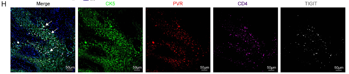
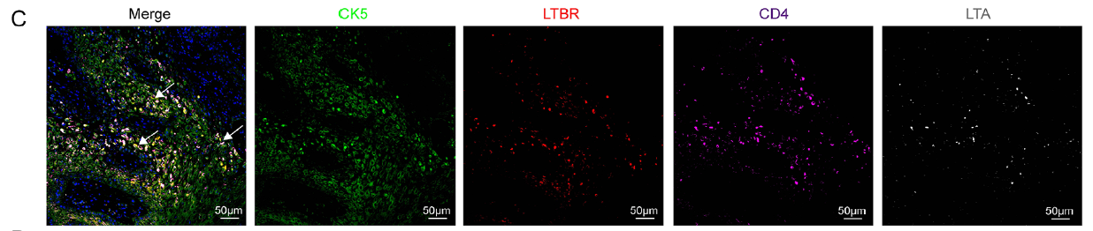
Plac1+ tumor cells recruit CD4+ T cells via the CXCL11/CXCR3 axis, then induce their differentiation into Tregs through the PVR/TIGIT pathway, constructing an immunosuppressive tumor microenvironment. Simultaneously, differentiated Tregs can reciprocally activate the PI3K/AKT signaling pathway in Plac1+ tumor cells via the LTA/LTBR axis, further enhancing their malignant phenotypes. This ultimately forms a Plac1+ tumor cell-Treg pro-tumorigenic positive feedback loop that continuously drives HNSCC progression.
III. Absin Products Providing Comprehensive Support for Reliable Experimental Validation
Multiple critical experimental steps in this study utilized Absin's high-quality products, including antibodies and mIHC kits. Their excellent specificity and sensitivity ensured accurate and reliable experimental results, becoming important contributors to the successful publication of the research findings. Below are the specific applications and roles of Absin products in this study:
Absin Products Used in This Study
| Catalog No. | Product Name | Applications in This Study |
|---|---|---|
| abs103873 | Rabbit anti-PLAC1 Polyclonal Antibody(C-term) |
|
| abs50012 | Four-Color Multiplex Immunofluorescence Staining Kit (Universal Secondary Antibody for Mouse/Rabbit) |
|
1. Absin Anti-Plac1 Antibody (abs103873)
Experimental Applications:
- Validation of CRISPR/Cas9 Plac1 knockout cell lines (Western blot)
- Immunohistochemical (IHC) detection of Plac1 in mouse tissues
Corresponding Figures: Figure S6A/B, Figure S7E/F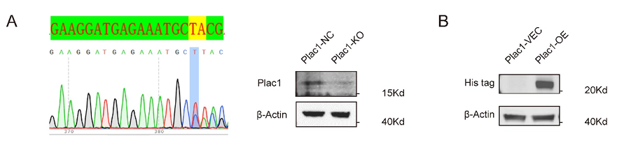

Product Role:
This antibody specifically recognizes Plac1 protein, successfully validating the construction efficiency of Plac1 knockout/overexpression cell lines in WB experiments, and clearly detecting Plac1 expression localization in mouse tissues via IHC. This established the cellular models and experimental foundation for subsequent functional studies of Plac1.
2. Absin PANO 4-plex IHC Kit (abs50012)
Experimental Applications:
Multiplex immunofluorescence (mIHC) staining of Plac1, Treg, and related molecules in human HNSCC tissues
Corresponding Figures: Figure 6C/D, Figure 6H, Figure 7C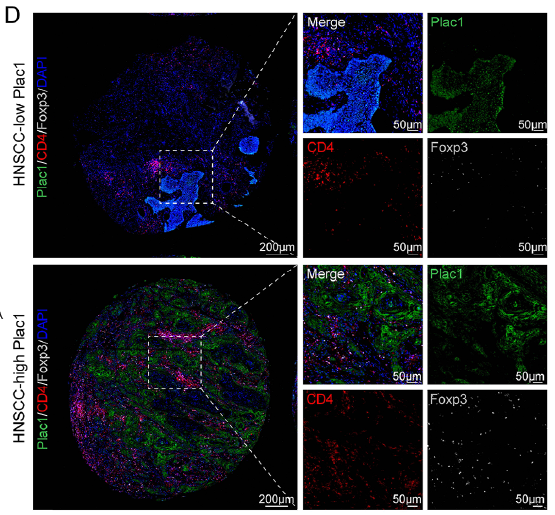
Product Role:
This kit supports synchronous fluorescent staining of multiple targets, enabling co-localization detection of Plac1 with CD4, Foxp3, PVR with TIGIT, and LTBR with LTA on the same tissue section. It clearly revealed the spatial interaction between Plac1+ tumor cells and Tregs and the binding patterns of key molecules, providing direct histological evidence for demonstrating the mutual regulation between Plac1+ tumor cells and Tregs.
IV. Research Significance and Future Perspectives
This study represents the first identification of Plac1 as an HNSCC-specific CTA, systematically elucidating its dual pro-tumorigenic mechanisms through cell-autonomous PI3K/AKT pathway regulation and construction of Treg-mediated immunosuppressive microenvironment with positive feedback loop formation. This not only enriches functional studies of CTAs in HNSCC but also provides new therapeutic targets—Plac1 and its associated regulatory pathways (CXCL11/CXCR3, PVR/TIGIT, LTA/LTBR) show potential as immunotherapy targets for HNSCC, offering novel strategies for addressing ICI resistance in HNSCC.
Absin remains committed to providing high-quality, high-specificity biological reagent products for life science research. The successful publication of this important research by Professor He Yue's team in a top-tier journal, supported by multiple Absin products, represents high recognition of Absin's product quality and technical services. Moving forward, Absin will continue to deepen its presence in the biological reagent field, continuously developing and launching more premium products to provide more comprehensive and reliable tool support for research in oncology, immunology, cell biology, and other fields, helping more researchers overcome scientific challenges and publish high-impact research achievements.
This article is based on the original publication in Advanced Science (DOI:10.1002/advs.202417312), interpreted and organized by AI. All original figures, data, and intellectual property rights belong to the original journal and research team. If any infringement exists, please contact us promptly for removal, and we will actively cooperate in handling such matters.
Contact Absin
Absin provides antibodies, proteins, ELISA kits, cell culture, detection kits, and other research reagents. If you have any product needs, please contact us.
| Absin Bioscience Inc. worldwide@absin.cn |
 Follow us on Facebook: Absin Bio Follow us on Facebook: Absin Bio |