- Cart 0
- English
How ECL Chemiluminescent Detection Kits Enhance Protein Detection Sensitivity and Reliability
March 05, 2026
Clicks:78
Western Blot, as the gold standard technique for protein detection, has its signal detection step directly determining experimental sensitivity and reproducibility. Traditional chromogenic methods (such as DAB, TMB), while operationally intuitive, offer limited sensitivity and are difficult to quantify. The emergence of ECL (Enhanced Chemiluminescence) technology has achieved orders-of-magnitude improvements in protein detection sensitivity through enzyme-catalyzed chemical reactions producing light signals. This article delves into the working principles, technical characteristics, and applications of ECL chemiluminescent detection kits in various experimental contexts.
What is an ECL Chemiluminescent Detection Kit?
An ECL chemiluminescent detection kit is a chemiluminescent detection system based on Horseradish Peroxidase (HRP) catalytic reactions. The kit typically comprises two core components: Solution A (luminol substrate solution) and Solution B (hydrogen peroxide/enhancer solution). When mixed in equal volumes, an oxidation reaction occurs under HRP catalysis, generating detectable chemiluminescent signals.
ECL kits are developed based on next-generation enhanced chemiluminescent substrates with optimized components. The product features low background and excellent stability. Catalyzed by horseradish peroxidase, the chemical reaction produces detectable light signals suitable for X-ray film exposure, direct chemiluminescence imager detection, or fluorescent CCD scanning.
According to detection sensitivity levels, ECL kits are typically classified into three tiers: Standard, Ultra-Sensitive, and Pico-Sensitive, suitable for detecting target proteins of different abundance levels.
Why Can ECL Achieve Ultra-High Sensitivity Detection?
Enzymatic Signal Amplification is the core mechanism underlying ECL's high sensitivity. A single HRP molecule can catalyze thousands of substrate molecules during the reaction, generating massive quantities of photons. This signal amplification effect enables detectable signals even from trace amounts of target protein, with detection sensitivity reaching picogram (pg) or even femtogram (fg) levels.
Low Chemiluminescent Background. Unlike fluorescent detection, chemiluminescence requires no external excitation light source, avoiding background interference caused by excitation light scattering. Luminescent signals are produced only at HRP-presenting locations, with signal-to-noise ratios significantly superior to chromogenic methods and certain fluorescent approaches.
Extended Signal Duration. ECL luminescence persists for extended periods, though fluorescence is strongest within the first 30 minutes following reaction initiation, gradually diminishing thereafter. This characteristic provides ample operational time windows while requiring experimenters to maximize utilization of this high-intensity "golden period" for film exposure or imaging.
Excellent Spectral Compatibility. ECL emission wavelengths match well with the sensitive spectral bands of X-ray films and CCD imaging systems, requiring no special filters or detection equipment—standard laboratory equipment suffices for signal acquisition.
How to Select Among Different Sensitivity Levels?
| ECL Type | Detection Sensitivity | Signal Duration | Antibody Dilution Ratio | Applicable Scenarios |
|---|---|---|---|---|
| Standard ECL | Mid-picogram | <12 hours | Primary: 1:1000-1:4000 Secondary: 1:1000-1:4000 |
Routine protein sample detection with high target abundance; moderate sensitivity with strong signals; suitable for most conventional Western Blot experiments |
| Ultra-Sensitive ECL | Low picogram | <12 hours | Primary: 1:1000-1:10000 Secondary: 1:2000-1:10000 |
Low target abundance with limited sample availability; cost-effective with high sensitivity; applicable to most Western Blot experiments; the laboratory workhorse product |
| Pico-Sensitive ECL | Low femtogram | <8 hours | Primary: 1:5000-1:100000 Secondary: 1:100000-1:500000 |
Trace protein detection with extremely low target abundance and precious samples; exceptionally strong sensitivity; ideal for trace protein detection; significantly reduces consumption of valuable antibodies |
Which Experimental Scenarios Can Be Applied?
Routine Western Blot Protein Detection represents the most fundamental application of ECL kits. This includes detecting target protein expression levels from cell lysates or tissue homogenates, validating gene knockout/overexpression effects, or comparing protein expression changes under different treatment conditions.
Protein-Protein Interaction Verification such as detection following Co-Immunoprecipitation (Co-IP). The high sensitivity of ECL facilitates detection of weak interactions or low-abundance protein complexes.
Post-Translational Modification Detection including phosphorylation, acetylation, ubiquitination, and other modified proteins. Since modified proteins typically constitute low proportions of total protein, high-sensitivity detection systems are required.
Subcellular Localization Studies where Western Blot is performed after fractionation of nuclei, cytoplasm, mitochondria, and other cellular components; ECL enables detection of marker proteins in each fraction.
Chemiluminescent ELISA Detection can employ ECL systems. Compared to traditional chromogenic methods, chemiluminescent ELISA offers wider linear range and higher sensitivity, suitable for quantitative detection of low-abundance cytokines.
Nucleic Acid Hybridization Detection such as signal detection for HRP-labeled probes in Southern Blot and Northern Blot applications.
How to Properly Use ECL Kits?
Working Solution Preparation:
Following conventional Western Blot procedures and secondary antibody incubation, prepare the chemiluminescent detection working solution during the final wash step according to membrane size. Typically mix 0.5 mL Solution A and 0.5 mL Solution B per 10 cm² membrane, equilibrate to room temperature after mixing. Immediate use is recommended; while usable after several hours at room temperature, sensitivity may be slightly reduced.
Membrane Processing:
Using flat forceps, remove the membrane and gently touch the lower edge to absorbent paper to remove excess liquid. Avoid complete membrane drying. Fully immerse the membrane in working solution (125 μL working solution per cm² membrane) to ensure complete contact. Incubate at room temperature for 2-3 minutes, then proceed immediately to film exposure.
Extended incubation times will not increase sensitivity and may sometimes cause abnormal band exposure. The luminescence process is fundamentally enzymatic; insufficient working solution hinders the reaction and causes uneven band exposure with significantly reduced sensitivity. To conserve reagents, membranes may be trimmed smaller, but do not reduce the working solution ratio.
Signal Detection:
Lift the membrane with forceps, drain excess working solution on filter paper, but do not wash away the working solution. Place plastic wrap larger than the membrane on the inner surface of an X-ray film cassette. Affix the blot to the plastic wrap, fold the wrap to completely enclose the blot, removing bubbles and wrinkles; trim excess wrap from edges. Absorb excess working solution with filter paper. Secure the wrap-covered blot in the cassette with tape, protein side facing upward.
In a darkroom, place an X-ray film on the wrapped membrane, close the cassette, and expose for 30 seconds to 1 minute. Develop and fix immediately; adjust exposure time for subsequent films based on intensity. Alternatively, proceed directly to chemiluminescence imager detection or fluorescent CCD scanning.
What Are the Critical Usage Considerations?
- Prevent Cross-Contamination Between Solutions A and B. When aspirating ECL Solutions A and B, pipette tips must be changed. Mutual contamination between Solutions A and B will cause gradual inactivation, affecting subsequent performance. Dedicated pipette tips or dispensers are recommended for each solution.
- Securely Cap Bottles Immediately After Use. Replace caps promptly after use to prevent inactivation. Solution B particularly, containing oxidizers, is susceptible to reduction and inactivation.
- Maximize the Golden 30 Minutes. While ECL luminescence persists for extended periods, fluorescence is strongest within the first 30 minutes following reaction initiation, gradually diminishing thereafter. Therefore, maximize utilization of this high-intensity period for film exposure.
- Safety Precautions. Both ECL Solutions A and B are harmful to humans; appropriate protective measures are required during operation. For safety and health, wear laboratory coats and disposable gloves.
- Membrane Selection. All reagents are compatible with both NC and PVDF membranes. PVDF membranes offer stronger protein binding capacity, suitable for low-abundance protein detection; NC membranes provide lower background, suitable for high-abundance proteins.
- Antibody Dilution Optimization. Different sensitivity-level ECL kits correspond to different recommended antibody dilution ratios. Refer to the recommended ranges for the corresponding level to avoid high background from excessive antibody concentration or weak signals from insufficient concentration.
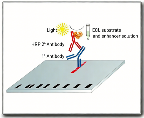
Schematic Diagram of ECL Chemiluminescent Detection Principle
Conclusion
ECL chemiluminescent detection kits provide high-sensitivity, low-background, and operationally convenient solutions for protein detection through enzyme-catalyzed chemiluminescent reactions. From routine protein expression to trace protein detection, from Western Blot to chemiluminescent ELISA, different sensitivity-level ECL kits satisfy diverse experimental requirements. Correctly mastering key points of working solution preparation, membrane processing, and signal acquisition, while optimizing antibody dilution ratios and exposure times, will significantly improve experimental success rates and data quality. With advancing imaging technologies, ECL chemiluminescent detection is progressively transitioning from traditional X-ray film to digital CCD imaging, providing more precise and quantitative analytical tools for protein research.
Contact Absin
Absin provides antibodies, proteins, ELISA kits, cell culture, detection kits, and other research reagents. If you have any product needs, please contact us.
| Absin Bioscience Inc. worldwide@absin.cn |
 Follow us on Facebook: Absin Bio Follow us on Facebook: Absin Bio |