- Cart 0
- English
Breaking the Oncolytic-Virotherapy Bottleneck in Pancreatic Cancer: Absin Uncovers the Monocyte–CAF–DC Immunosuppressive Axis, Empowering a Novel Combination Strategy
December 01, 2025
Clicks:76
Reference: Li F, et al., J Immunother Cancer 2025
Title: Oncolytic virus-induced IL-1β⁺ monocyte–IL-6⁺ CAF axis suppresses dendritic cell-mediated antitumor immunity in pancreatic cancer
Pancreatic ductal adenocarcinoma (PDAC), one of the most lethal malignancies, exhibits a 5-year survival rate <10 %. Its dense, highly immunosuppressive tumor microenvironment (TME) severely limits the efficacy of immunotherapy. Although oncolytic virus (OV) therapy has shown potential to reactivate antitumor immunity, how stromal components modulate OV efficacy remains poorly understood.
A recent breakthrough in the Journal for ImmunoTherapy of Cancer identifies an OV-induced “IL-1β⁺ monocyte–IL-6⁺ CAF axis” that cripples dendritic cell (DC)-mediated antitumor immunity. Using Absin’s TSA multiplex immunofluorescence kits, the authors achieved high-resolution spatial mapping of immune–stromal interactions, providing critical support for dissecting the immunosuppressive network.
I. Experimental Strategy: Dissecting OV Resistance Layer-by-Layer
Clinical problem: PDAC TME is rich in cancer-associated fibroblasts (CAFs) that create an immune barrier and blunt OV efficacy. The team interrogated the tripartite cross-talk among virus, immune cells, and stroma to uncover resistance mechanisms.
Step-wise validation:
- Evaluate antitumor activity of engineered HSV-OX40L/IL-12 and its impact on immune compartments;
- Trace the cellular and cytokine drivers that impair DC maturation;
- Define the signaling cascade: monocyte-IL-1β → CAF-IL-6 → DC paralysis;
- Develop and validate combination regimens in murine models and patient-derived specimens.
Technical arsenal: single-cell RNA-seq, flow cytometry, multiplex immunofluorescence, ELISA, and functional assays.
II. Key Findings: Unlocking a Novel Immunosuppressive Axis and a Route to Combination Therapy
(1) Discovery of the IL-1β⁺ monocyte–IL-6⁺ CAF axis driving OV resistance
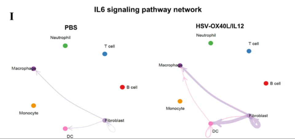
OV infection releases DAMPs/PAMPs that activate monocytes to secrete IL-1β; IL-1β in turn stimulates CAFs to produce IL-6, generating an IL-6-high milieu that directly blocks DC maturation and co-stimulation, ultimately crippling T-cell-mediated immunity. Targeting this “monocyte–CAF–DC” axis offers a new strategy to overcome OV resistance.
(2) Next-gen OV + CD40 agonist + FLT3L achieves triple synergy
An HSV vector encoding FLT3L, OX40L, and IL-12 was combined with CD40 agonistic antibody:
- Restored DC activation (high CD80/CD86) and antigen-presenting capacity;
- Elicited potent tumor-specific T-cell responses with 40 % complete regression;
- Cured mice developed long-term immune memory against re-challenge.
(3) Tumor-draining lymph nodes (TDLNs) are essential for immune activation
Surgical removal of TDLNs abolished the therapeutic effect, confirming TDLNs as the anatomical hub for DC priming of T cells and guiding treatment optimization.
III. Powered by Absin: TSA Multiplex Immunofluorescence Illuminates Immune–Stromal Interactions
(1) Core product & samples
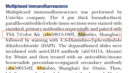
TSA 7-Color Kit (cat# abs50015-100T)
| Parameter | Details |
| Species | C57BL/6J mice + Batf3⁻/⁻ knockout |
| Tumor models | ① Murine Panc02 cell line ② GEMM KPC autochthonous PDAC line |
| Tissue | Subcutaneous tumors (FFPE) |
| Treatment arms | PBS → HSV-OX40L/IL-12 → HSV-OX40L/IL-12/FLT3L+αCD40 |
| Controls | Isotype IgG, Batf3⁻/⁻ (cDC1-deficient), CD8⁺ T-cell depletion, TDLN resection |
(2) Application in the study
Multiplex immunofluorescence was employed to visualize immune-cell infiltration and spatial interactions after therapy:
| Item | Protocol | Purpose |
| Technology | 7-color TSA multiplex IHC | Simultaneous detection of B cells, T cells, DCs plus nuclear counterstain on a single section |
| Kit | Absin TSA 7-Color Kit (abs50015-100T) | Tyramide signal amplification with cyclic stripping/re-staining ensures sensitivity and specificity |
| Antibody/fluor order | 1. CD19 → TSA 520 (green) 2. CD3 → TSA 650 (far-red) 3. CD11c → TSA 570 (yellow) 4. DAPI (blue, nuclei) |
Specific antibody-fluorophore pairs identify B cells, T cells, and DCs; spatial relationships resolved |
| Section type | 4 µm FFPE sections of murine subcutaneous Panc02/KPC tumors | Collected day 19 post-treatment; clinically relevant format to assess immune remodeling |
| Imaging & analysis | Pannoramic MIDI II whole-slide scanner + Indica HALO software | Low-power (200 µm scale bar) overview and high-power (50 µm) quantification of tertiary lymphoid structures |
(3) Product advantages & mechanistic insights
1. Spatial phenotyping
mIHC revealed that only the triple-combination group (HSV-OX40L/IL-12/FLT3L + αCD40) generated CD11c⁺ DC–CD3⁺ T–CD19⁺ B cell clusters within tumor parenchyma, indicative of functional tertiary lymphoid structures (TLS).
2. Functional correlation
- Flow cytometry: highest GzmB⁺ IFN-γ⁺ CD8⁺ T-cell fraction
- Survival: 40 % complete responses; re-challenge 100 % rejected
- Mechanism: DC co-stimulatory molecules (CD80/86) and IL-12 production significantly elevated
→ mIHC “spatial clusters” directly aligned with systemic immune protection.
3. Mechanistic conclusion
OV → DAMP/PAMP → monocyte IL-1β → CAF IL-6 → DC paralysis;
IL-6 blockade or concurrent CD40 + FLT3 agonism restores DC function, and mIHC visually documents DC re-aggregation and formation of a functional immune microenvironment.

This study unveils a previously unrecognized resistance mechanism to oncolytic virotherapy in pancreatic cancer and delivers a mechanism-based combination regimen, offering new hope for this recalcitrant malignancy. Powered by high-sensitivity, high-specificity Absin TSA multiplex immunofluorescence kits, the team successfully visualized the immune–stromal interaction network, providing critical technical support for mechanistic validation.
Moving forward, Absin will continue to develop premium tools tailored to tumor immunology and cellular-interaction studies, partnering with researchers to conquer the toughest challenges in cancer therapy and to accelerate the innovation and translation of immunotherapies.Related Products
| Cat. No. | Product Name | Size |
|---|---|---|
| abs50086 | 2-Color Multiplex Immunofluorescence Kit (anti-rabbit secondary) | 100T |
| abs50087 | 2-Color Multiplex Immunofluorescence Kit (mouse/rabbit universal secondary) | 100T |
| abs50088 | 3-Color Multiplex Immunofluorescence Kit (anti-rabbit secondary) | 100T |
| abs50089 | 3-Color Multiplex Immunofluorescence Kit (mouse/rabbit universal secondary) | 100T |
| abs50012 | 4-Color Multiplex Immunofluorescence Kit (mouse/rabbit universal secondary) | 20T/50T/100T |
| abs50168 | 4-Color Multiplex Immunofluorescence Kit B (anti-rabbit secondary) | 20T/50T/100T |
| abs50013 | 5-Color Multiplex Immunofluorescence Kit (mouse/rabbit universal secondary) | 20T/50T/100T |
| abs50029 | 5-Color Multiplex Immunofluorescence Kit (anti-rabbit secondary) | 20T/50T/100T |
| abs50014 | 6-Color Multiplex Immunofluorescence Kit (mouse/rabbit universal secondary) | 20T/50T/100T |
| abs50030 | 6-Color Multiplex Immunofluorescence Kit (anti-rabbit secondary) | 20T/50T/100T |
| abs50048 | 6-Color Multiplex Immunofluorescence Kit (plus) (anti-rabbit secondary) | 20T/50T/100T |
| abs50049 | 6-Color Multiplex Immunofluorescence Kit (plus) (mouse/rabbit universal secondary) | 20T/50T/100T |
| abs50015 | 7-Color Multiplex Immunofluorescence Kit (mouse/rabbit universal secondary) | 20T/50T/100T |
| abs50031 | 7-Color Multiplex Immunofluorescence Kit (anti-rabbit secondary) | 20T/50T/100T |
| abs50037 | 7-Color Multiplex Immunofluorescence Kit (plus) (mouse/rabbit universal secondary) | 20T/50T/100T |
| abs50038 | 7-Color Multiplex Immunofluorescence Kit (plus) (anti-rabbit secondary) | 20T/50T/100T |
| abs50165 | 7-Color Multiplex Immunofluorescence Kit (DyLight 770 enhanced) (anti-rabbit secondary) | 20T/50T/100T |
| abs50166 | 7-Color Multiplex Immunofluorescence Kit (DyLight 770 enhanced) (mouse/rabbit universal secondary) | 20T/50T/100T |
| abs50018 | 10-Color Multiplex Immunofluorescence Kit | 100T |
| abs50083 | Lung-Cancer Tumor-Microenvironment Multiplex Immunofluorescence Kit (I) | 20T |
| abs50084 | Lung-Cancer Tumor-Microenvironment Multiplex Immunofluorescence Kit (II) | 20T |
Contact Absin
Absin provides antibodies, proteins, ELISA kits, cell culture, detection kits, and other research reagents. If you have any product needs, please contact us.
| Absin Bioscience Inc. worldwide@absin.cn |
 Follow us on Facebook: Absin Bio Follow us on Facebook: Absin Bio |