- Cart 0
- English
[IF 20.4] Breakthrough! 3D Reconstruction of Human CS9 Embryo with Absin Tools Cracks the Black Box of Early Development
December 01, 2025
Clicks:77
A landmark study recently published in Cell Stem Cell presents the first 3-D reconstruction of a human Carnegie Stage 9 (CS9) embryo using spatial transcriptomics, offering an unprecedented snapshot of early body-plan formation during organogenesis. As a dedicated supplier of research tools, Absin products played a pivotal role in this cutting-edge work, enabling precise validation of cell positioning and function and providing robust technical support for embryonic-development research.
Title: 3D reconstruction of a human Carnegie stage 9 embryo provides a snapshot of early body-plan formation
Journal: Cell Stem Cell (IF 20.4)
DOI: https://doi.org/10.1016/j.stem.2025.04.007
Key Reagents: 7-Color Multiplex Immunofluorescence Kit (abs50015), Antibody Stripping Buffer (abs994)
I. Research Highlights: Decoding the “Critical Window” of Embryogenesis
CS9 embryos (19–21 days post-fertilization) mark the transition from late gastrulation to early organogenesis, representing the “golden period” when the heart, neural tube, gut and other vital organs are initiated. Ethical constraints and sample scarcity have long kept this stage a “black box” in human developmental biology.
Key breakthroughs:
- First 3-D spatial transcriptome atlas of a CS9 embryo, identifying 27 major cell populations covering brain, spine, primitive gut, somites, etc.
- Dual origin of the hindbrain (neuro-ectoderm + neuromesodermal progenitors, NMPs) and the underlying regulatory mechanism.
- Mapping the mid-hindbrain organizer (IsO) and anterior intestinal portal (AIP), confirming AIP’s organizer role in heart development.
- Early aorta formation and presence of primordial germ cells (PGCs) in the AGM region, offering new clues to hematopoietic stem-cell origins.
II. Experimental Strategy: Spatial Transcriptomics + 3-D Modeling for Molecular & Structural Insights
The authors applied a closed-loop workflow—“section sequencing → data integration → 3-D reconstruction → functional validation”—to decipher developmental puzzles step-by-step:
- Sample preparation: Normal karyotype CS9 embryo embedded in OCT; 75 serial 10-µm transverse sections collected.
- Spatial transcriptomics: Stereo-seq captured gene-expression information; unsupervised clustering identified cell types.
- 3-D reconstruction: PASTE algorithm aligned all sections and integrated spatial coordinates with expression data to generate a visual 3-D model.
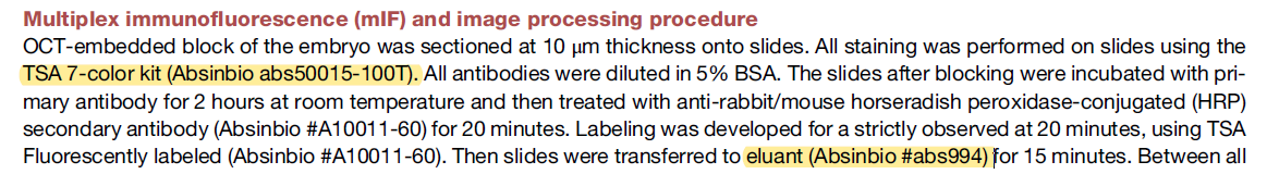
- Functional validation: Multiplex immunofluorescence (mIF) confirmed localization and marker expression of key populations (e.g., hemogenic endothelium, PGCs), providing morphological evidence for transcriptomic data.
- Cross-species & in-vitro comparison: CS9 data were compared with mouse, monkey embryos and human pluripotent-stem-cell-derived models to verify reliability.
III. Absin Product Application: Core Tools for Multiplex Immunofluorescence
For functional validation the team selected three Absin reagents and successfully accomplished precise localization and visualization of cell markers (Fig. 6F):
| Absin Product | Cat. No. | Key Function |
|---|---|---|
| 7-Color Multiplex Immunofluorescence Kit | abs50015 | Simultaneous staining of CD31, CD34, CDH5 (hemogenic endothelium) and BLIMP1, TFAP2C (PGCs); clear spatial discrimination of cell populations |
| Antibody Stripping Buffer | abs994 | Enables multiple antigen labeling on the same section, solving co-localization challenges and improving staining efficiency & accuracy |

Figure: Multiplex IF of the CS9 AGM region (corresponds to Fig. 6F in the original paper)
Staining targets:
Hemogenic endothelium (CD31⁺, CD34⁺, CDH5⁺) and primordial germ cells (BLIMP1⁺, TFAP2C⁺) in the AGM.
Product value:
Absin’s TSA-based kit ensured clear separation of five markers; the stripping buffer supported multi-round labeling, while HRP-conjugated secondary antibodies guaranteed signal intensity and specificity. The final images revealed hemogenic endothelium surrounding the hindgut and PGCs localized at the AT-AGM transition, directly validating the spatial-transcriptomic cell-type assignments.
IV. Product Core Value: Reliable Support for “Spatial + Functional” Validation
- High specificity: TSA technology minimizes background, enabling accurate detection of low-abundance markers in complex embryonic tissues.
- Multicolor compatibility: 7-color kit meets co-localization needs without spectral overlap, ideal for cell-type validation.
- Workflow integration: Stripping buffer and staining kit work in tandem to support multi-round labeling, boosting efficiency and conserving precious human-embryo samples.
V. Summary & Outlook
This study fills a critical knowledge gap in early human embryogenesis and provides an invaluable reference atlas for developmental biology, regenerative medicine and congenital-disease research. As a research-tool provider, Absin remains committed to delivering precise and reliable reagents—from multiplex immunostaining to signal-amplification systems—to empower cutting-edge science.
Products used in the paper:
| Cat. No. | Name | Size |
|---|---|---|
| abs50015 | 7-Color Multiplex Immunofluorescence Kit (mouse/rabbit universal secondary) | 20T/50T/100T |
| abs994 | Antibody Stripping Buffer (for mIHC) | 30 mL |
More multiplex immunofluorescence kits
| Cat. No. | Product Name | Size |
|---|---|---|
| abs50086 | 2-Color Multiplex Immunofluorescence Kit (anti-rabbit secondary) | 100T |
| abs50087 | 2-Color Multiplex Immunofluorescence Kit (mouse/rabbit universal secondary) | 100T |
| abs50088 | 3-Color Multiplex Immunofluorescence Kit (anti-rabbit secondary) | 100T |
| abs50089 | 3-Color Multiplex Immunofluorescence Kit (mouse/rabbit universal secondary) | 100T |
| abs50012 | 4-Color Multiplex Immunofluorescence Kit (mouse/rabbit universal secondary) | 20T/50T/100T |
| abs50168 | 4-Color Multiplex Immunofluorescence Kit B (anti-rabbit secondary) | 20T/50T/100T |
| abs50013 | 5-Color Multiplex Immunofluorescence Kit (mouse/rabbit universal secondary) | 20T/50T/100T |
| abs50029 | 5-Color Multiplex Immunofluorescence Kit (anti-rabbit secondary) | 20T/50T/100T |
| abs50014 | 6-Color Multiplex Immunofluorescence Kit (mouse/rabbit universal secondary) | 20T/50T/100T |
| abs50030 | 6-Color Multiplex Immunofluorescence Kit (anti-rabbit secondary) | 20T/50T/100T |
| abs50048 | 6-Color Multiplex Immunofluorescence Kit (plus) (anti-rabbit secondary) | 20T/50T/100T |
| abs50049 | 6-Color Multiplex Immunofluorescence Kit (plus) (mouse/rabbit universal secondary) | 20T/50T/100T |
| abs50015 | 7-Color Multiplex Immunofluorescence Kit (mouse/rabbit universal secondary) | 20T/50T/100T |
| abs50031 | 7-Color Multiplex Immunofluorescence Kit (anti-rabbit secondary) | 20T/50T/100T |
| abs50037 | 7-Color Multiplex Immunofluorescence Kit (plus) (mouse/rabbit universal secondary) | 20T/50T/100T |
| abs50038 | 7-Color Multiplex Immunofluorescence Kit (plus) (anti-rabbit secondary) | 20T/50T/100T |
| abs50165 | 7-Color Multiplex Immunofluorescence Kit (DyLight 770 enhanced) (anti-rabbit secondary) | 20T/50T/100T |
| abs50166 | 7-Color Multiplex Immunofluorescence Kit (DyLight 770 enhanced) (mouse/rabbit universal secondary) | 20T/50T/100T |
| abs50018 | 10-Color Multiplex Immunofluorescence Kit | 100T |
| abs50083 | Lung-Cancer Tumor-Microenvironment Multiplex Immunofluorescence Kit (I) | 20T |
| abs50084 | Lung-Cancer Tumor-Microenvironment Multiplex Immunofluorescence Kit (II) | 20T |
【Disclaimer】 This article is compiled from the original publication Cell Stem Cell (DOI: 10.1016/j.stem.2025.04.007) by AI. All images and data rights belong to the original journal and authors. If any infringement is found, please contact us for prompt removal.
Contact Absin
Absin provides antibodies, proteins, ELISA kits, cell culture, detection kits, and other research reagents. If you have any product needs, please contact us.
| Absin Bioscience Inc. worldwide@absin.cn |
 Follow us on Facebook: Absin Bio Follow us on Facebook: Absin Bio |