- Cart 0
- English
Three-step construction of atherosclerotic cell model: a complete strategy for ox-LDL-induced injury/foam/calcification
August 28, 2025
Clicks:567
Atherosclerosis (AS) is a chronic cardiovascular disease that endangers human health and is one of the most common causes of death in the elderly. The main pathological feature of AS is lipid deposition in some arteries, accompanied by smooth muscle cells (SMCs) and fibrous matrix hyperplasia, which gradually develops into AS plaques.
Vascular calcification (VC) is a pathological phenomenon characterized by calcium phosphate deposition on the walls of arteries and veins. Patients with atherosclerosis (AS), diabetes mellitus and chronic kidney disease all have a great risk of developing VC.
The accumulation of oxidized low-density lipoprotein (ox-LDL) in blood vessel walls plays a key role in the development of AS and VC. Studies have shown that high expression of ox-LDL has been detected in the serum of patients with AS cerebral infarction, and it is believed that it can accelerate the formation of carotid plaque and promote the shedding of unstable plaque, which is an independent risk factor for cerebrovascular disease. Therefore, ox-LDL, AS a key pathogenic factor, is an effective model of AS.
The pathogenesis of AS is complex, and the processes of cell injury, foaming, calcification and other processes are intertwined and influence each other. There is no absolute and invariable sequence, but it is generally believed that endothelial cell injury is the initial step of atherosclerosis. Under the action of many factors such as hyperlipidemia, hypertension, smoking, diabetes, etc., vascular endothelial cells are damaged, their permeability increases, and lipids and inflammatory cells in blood can enter the subendothelium.
After endothelial cell injury, lipids such as low-density lipoprotein cholesterol (LDL-C) in blood enter the subendothelial space and are oxidatively modified to form ox-LDL. ox-LDL is phagocytosed by macrophages, forming a large number of lipid droplets in the cell, and the macrophages are converted into foam cells. Foam cell aggregation is an early lesion of atherosclerosis. With the development of atherosclerotic lesions, smooth muscle cells proliferate, migrate and undergo phenotypic transformation at the lesion site, and at the same time produce a large amount of extracellular matrix. In this process, local inflammatory reaction, cell death, etc. release some pro-calcification factors, such as alkaline phosphatase, etc., which promote the deposition of calcium salts on the blood vessel wall and lead to blood vessel calcification. Calcification usually occurs after the formation of atherosclerotic plaque, which makes the blood vessel wall hard and brittle, and further aggravates the vascular disease.
1. ox-LDL induced HUVECs injury model of human umbilical vein endothelial cells
01 Experimental cells
Human umbilical vein endothelial cells HUVECs (purchased from American Type Culture Collection, ATCC)
02 Method of inducing endothelial cell injury
① After HUVECs were inoculated, they were pretreated with different concentrations of PFE (1.5, 3.0, 7.5 μg/mL) for 2 h (Note: PFE is a flavonoid extracted from coriander seeds).
② ox-LDL stimulation: Remove the pretreated medium and add fresh medium containing 100μg/mL ox-LDL (abs47014903). The culture was continued for 24 hours to induce endothelial injury.
This model induces endothelial inflammation and adhesion molecule expression through ox-LDL (Fig. 1), simulating early endothelial injury in atherosclerosis, and is suitable for the mechanism study of anti-atherosclerotic drugs (such as PFE).
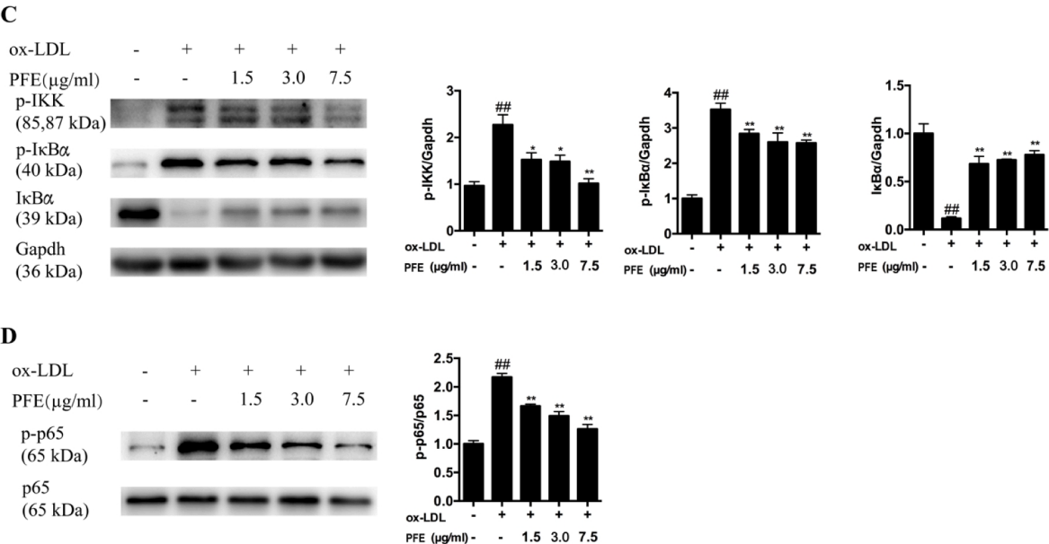
Fig. 1 PFE inhibits ox-LDL-stimulated inflammation in HUVECs by inhibiting the NF-kB pathway
2. ox-LDL-induced macrophage RAW 264.7 foaming model
01 Experimental cells
Macrophage cell line RAW 264.7 (purchased from American Type Culture Collection, ATCC)
02 Induced foaming method
① The experimental group was first pretreated with PFE (flavonoids extracted from coriander seeds) (1.5, 3.0, 7.5 μg/mL) for 2 hours. No PFE was added to the control group.
② ox-LDL stimulation: ox-LDL (abs47014903) was added to a final concentration of 100 μg/mL, and the cells were incubated for 24 hours. Detection index: oil red O (abs42024259) staining to observe lipid accumulation (degree of foaming). RT-PCR/Western blot detected gene expression related to cholesterol influx (LOX-1, CXCL16) and efflux (ABCA1, ABCG1, SR-B1) (Fig. 2).
This model successfully induced the transformation of RAW 264.7 macrophages into foam cells by ox-LDL, and confirmed that the drug PFE promoted cholesterol efflux and inhibited foaming through the PPARγ-ABCA1/ABCG1 pathway.
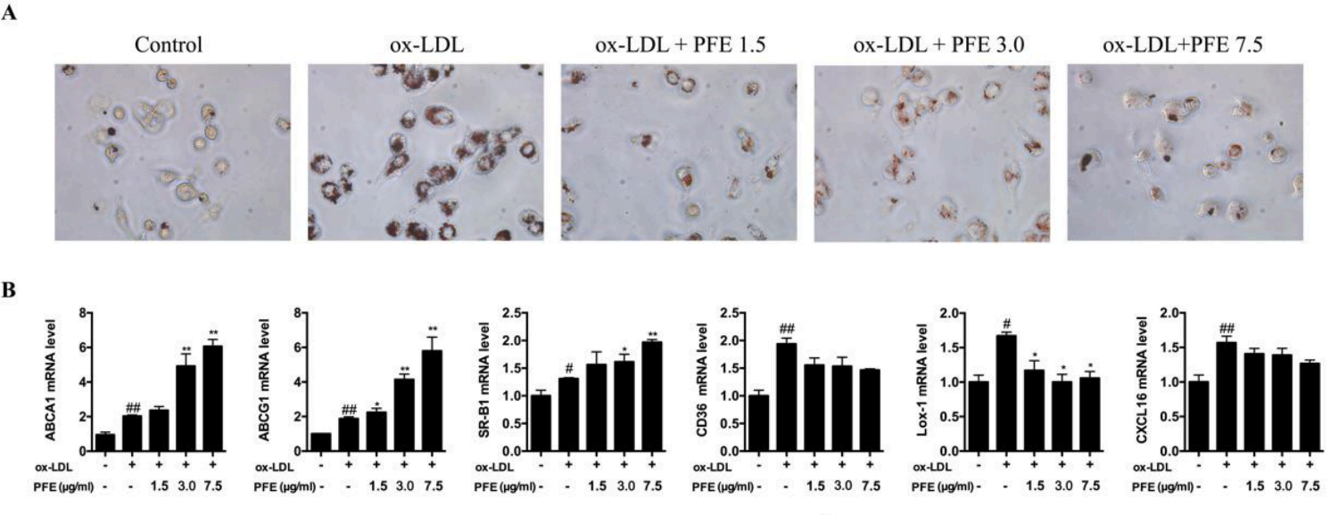
Fig. 2 PFE improves lipid accumulation in foam cells (representative images of foam cells stained with oil red O)
3. ox-LDL-induced smooth muscle cell SMCs calcification model
01 Experimental cells
Human aortic vascular smooth muscle cells HA-SMCs (purchased from CTCC cell bank, China)
02 Calcification modeling method
① HA-SMCs were divided into control group and calcification model group, and the cells after passage were starved for 24 hours, and the control group was replaced with complete medium;
② The calcification medium of the model was changed: 10mmol/L β-glycerophosphate (abs812987) + 50μg/mL L-ascorbic acid (abs811910) + 100nmol/L dexamethasone (abs813595); And ox-LDL (abs47014903) at a concentration of 50 μg/mL was added to induce cells for 14 days;
③ The results of alizarin red S (abs42012987) staining showed that the calcification model group had obvious orange-red calcification nodules compared with the control group, and the calcification model was successfully established.
This model establishes an SMCs calcification model by screening the optimal induction concentration of ox-LDL (Figure 3), which is suitable for studying the effect of specific anti-inflammatory factors (such as IL-37) on the calcification process of this model and its molecular mechanism.

Figure 3 Alizarin red calcification staining
In the long journey of atherosclerosis research, ox-LDL is undoubtedly a "golden key" in our hands. Through the above-mentioned well-designed cell model construction method, we can deeply explore its mystery in disease progression. As a solid backing for your scientific research journey, Absin provides various high-quality reagents and products that will help you ride the wind and waves in the field of atherosclerosis research and continuously achieve new breakthroughs and results.
References:
[1]Liu J, Zhang W, Li Y, Li X, Li Y, Guo F. Flavonoids extract from the seeds of Psoralea corylifolia L. (PFE) alleviates atherosclerosis in high-fat diet-induced LDLR-/- mice. Phytomedicine. 2022;98:153983.
doi:10.1016/j.phymed.2022.153983
[2]Machenyue. IL-37 inhibits calcification of human aortic vascular smooth muscle cells and is involved in inhibiting their osteogenic transformation [D]. Anhui Medical University, 2023.
Atherosclerosis Research Product Recommendation
|
Item number |
Product Name |
Specifications |
|
abs47014899 |
Human very low density lipoprotein |
2mg |
|
abs47014900 |
Human low density lipoprotein |
2mg |
|
abs47014901 |
Red fluorescent labeling of human low density lipoprotein |
500μg |
|
abs47014902 |
Green fluorescent labeling of human low density lipoprotein |
500μg |
|
abs47014903 |
Human oxidized low density lipoprotein |
2mg |
|
abs47014904 |
Human hyperoxidized low density lipoprotein |
2mg |
|
abs47014905 |
Human Red Fluorescent Labeled Oxidized Low Density Lipoprotein |
500μg |
|
abs47014906 |
Human acetylated low density lipoprotein |
2mg |
|
abs47014907 |
Red fluorescent labeling of human acetylated low density lipoprotein |
500μg |
|
abs47014908 |
Green fluorescent labeling of human acetylated low density lipoprotein |
500μg |
|
abs42024259 |
Oil Red O |
100g |
|
abs42018484 |
Philepine |
1mg |
|
abs812987 |
β-glycerophosphate |
1g |
|
abs811910 |
L-ascorbic acid |
500mg |
|
abs813595 |
Dexamethasone |
500mg |
|
abs580113 |
Low density lipoprotein detection kit |
96T |
|
abs580112 |
High density lipoprotein detection kit |
96T |
Absin provides antibodies, proteins, ELISA kits, cell culture, detection kits, and other research reagents. If you have any product needs, please contact us.
|
Absin Bioscience Inc. Email: worldwide@absin.cn |
 Follow us on Facebook: Absin Bio Follow us on Facebook: Absin Bio |