- Cart 0
- English
Is microglia research difficult? This guide unlocks key technical solutions
August 22, 2025
Clicks:98
As the resident immune cells of the central nervous system, microglia play an important role in immune surveillance, immune response and development and maintenance of the nervous system. Microglia are highly heterogeneous and sensitive. At present, the research on its complex regulatory mechanism and diverse functions has become an important direction in the field of brain science.
In vitro research: 4 types of cell model protocols
1. Primary cells
Applicable scenarios: research on mechanisms that need to retain in vivo characteristics to the greatest extent;
Breakthrough point: human/mouse brain tissue sorting technology (retaining original biological characteristics);
Precautions: Strictly aseptic operation to avoid phenotypic drift caused by long culture period.
2. Immortalized cell lines
Recommended cells: such as human HMC3, HMO6 and murine BV-2, N9, etc.;
Core advantages: low cost, short cycle, suitable for high-throughput drug screening;
Limitation suggests that long-term passage should be alert to genetic variation.
3. Inducing microglia-like cells
Technological breakthrough: iPSC differentiation technology + beta amyloid stimulation (abs45128173) = successfully simulated disease-related phenotype (DAM);
Application value: a golden tool for the mechanism study of genetic diseases.
4. Brain organoids
Cutting-edge scheme: PU.1 overexpression induces cortical organoids to generate functional microglia, and further xenotransplants organoid models into mice. Vascularized and remodeled organoids provide close nutrition and signal support in vivo for microglia development.
Advantages of the model: It studies the interaction between cells under three-dimensional structures, provides a physiological environment-like research platform, and is suitable for disease model construction.
In vivo research: 3 major animal model schemes
1. Drug depletion model [1]
The CSF1R small molecule inhibitor PLX5622 (abs823427) relies on the advantage of high penetration of the blood-brain barrier to target CSF1R for efficient clearance. Among them, PLX5622 can achieve 80% cell clearance rate after 3 days of administration, and the clearance effect can be maintained for up to 6 months. It has both high specificity and low inflammation risk, and has become the current gold standard for microglia depletion in mice.
2. Aging research model [2]
3-DR model: PLX5622 (abs823427) three rounds of depletion → directed simulated aging state
Mechanism of aging: three rounds of forced proliferation (> 20 divisions) → telomere loss/DNA damage accumulation → permanent aging
First round of drug administration --PLX5622 treatment for 7 days→ Microglial depletion >90% → Drug withdrawal for 7 days followed by repopulation → Second round of drug administration → Redepletion and repopulation → Third round of drug administration → Induction of cellular replicative senescence
Compared with natural aging, the cycle is shortened by 80%, avoiding the interference of multicellular synchronous aging.
3. Transgenic model
Microglia gene editing technology mainly includes Cre-loxP recombinase system, CRISPR/Cas gene editing technology and virus transduction technology.
|
Method |
advantage |
shortcoming |
|
Cre-loxP recombinase system |
Selective knockout of specific genes; Highly spatiotemporally specific, regulating specific developmental stages and cell types. |
Construction systems depending on specific promoters and target genes; The editing range is limited and only targets genes between loxP sites. |
|
CRISPR/Cas gene editing
|
Efficient editing ability, accurate editing of multiple genes at the same time. |
PAM sequence dependence restricts targeted editing range; Potential off-target risk triggers non-specific editing. |
|
Viral transduction |
Efficient transduction of large fragments of genes |
The expression time of foreign genes transduced by some viral vectors is insufficient. |
Microglia in vivo imaging technology
With the advantages of high tissue penetration, low background interference, and high spatiotemporal resolution, near-infrared region II (NIR-II, 1000-1700 nm) cyanine dyes provide breakthrough tools for microglia imaging in vivo.
Targeted Probe NIR-II Imaging
Probe design: Cyanine dyes such as ICG (abs816408), IR-820 (abs825392), etc. are coupled to ligands (such as antibodies, polypeptides) targeting microglia surface markers such as TREM2 (abs05663), CSF1R (abs06312), etc. to construct specific probes (such as HSA-ICG-iRGD in similar literature, Figure 1).
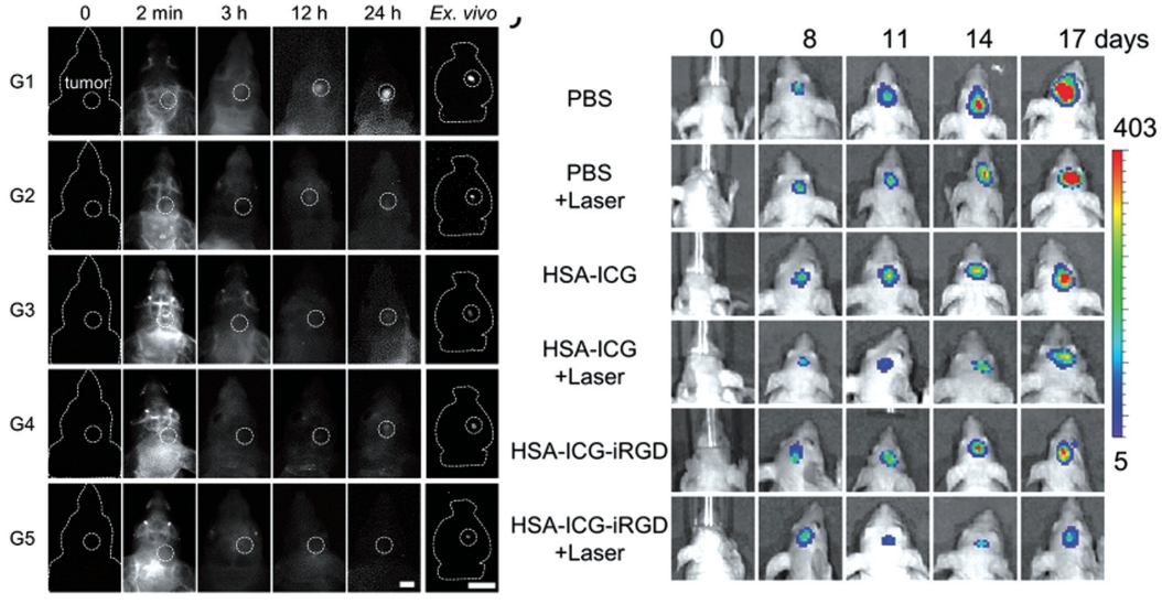
Fig. 1 Schematic diagram of HSA-ICG-iRGD actively targeting NIR-Ⅱ imaging and photothermal therapy in orthotopic glioma mice [3]
Imaging Advantages:
① The penetration depth reaches centimeter level (the penetration depth of brain tissue is increased by 3-5 times) to overcome the occlusion of the skull;
② Signal-to-noise ratio (SBR) > 6 (literature cases up to 6.85), clearly distinguishing activated/resting microglia;
③ Dynamically monitor the migration of microglia (such as the process of aggregation to Aβ plaques).
Application scenario: Real-time tracking of Aβ phagocytosis by microglia in Alzheimer's disease model.
From precise clearance, dynamic imaging to disease modeling, the bottleneck of microglia research is being systematically broken through! Whether it is the gold standard of three-day efficient depletion of PLX5622, the in vivo monitoring ability of NIR-II probe penetrating the skull, or the pathological model of iPSC differentiation + organoid construction, these technological innovations are pushing the research of neurological disease mechanisms into a new dimension.
References:
[1] Boland R, Kokiko-Cochran ON. Deplete and repeat: Microglial CSF1R inhibition and traumatic brain injury. Front Cell Neurosci. 2024,18:1352790.
[2] Li X, Li Y, Jin Y, et al. Transcriptional and epigenetic decoding of the microglial aging process.Nat Aging. 2023,3(10):1288~1311.
[3] Wu Y Y, Hu D H, Gao D Y, et al. Miniature NIR-Ⅱ nanoprobes for active-targeted phototheranostics of brain tumors[J]. Advanced Healthcare Materials, 2022, 11(23): e2202379.
Product recommendations in this issue:
|
Item number |
Product name |
Specifications |
|
abs823427 |
PLX5622 |
10 mg |
|
abs816408 |
Indocyanine Green (ICG) |
500 mg |
|
abs825392 |
IR-820 |
100 mg |
|
abs47047387 |
Human serum albumin (HSA) |
1g |
|
abs47048121 |
iRGD peptide |
1g |
|
abs45128173 |
Amyloid β-Protein (1-42) |
1mg |
|
abs44056601 |
Amyloid β-Protein (1-40) |
1mg |
|
abs45151799 |
Amyloid β-Protein (25-35) |
1mg |
|
abs05663 |
Recombinant Human TREM2 Protein(His Tag) |
100 μg |
|
abs06312 |
Recombinant Human CSF1R Protein(C-10His) |
100 μg |
|
abs90051 |
Organotial animal brain organoid medium |
100mL |
|
abs90050 |
Organotial animal brain organoid culture medium kit |
1kit |
Recommended products for induction of neurological disease models:
|
Item number |
Product Name |
Disease model |
|
abs42024900 |
Whooping cough toxin |
EAE model immune enhancer |
|
abs815889 |
MOG(35-55) |
EAE model immunogen, dominated by T cells, and mild demyelination. |
|
abs05465 |
MOG(1-125) |
EAE model immunogen, demyelinating + antibody-mediated injury (B cell involvement). |
|
abs9270 |
Freund's complete adjuvant |
Immune adjuvant, mixed with antigen and emulsified for use. |
|
abs814897 |
MPTP HCl |
Inducible Parkinson's model |
|
abs42070403 |
6-hydroxydopamine hydrobromide |
Inducible Parkinson's model |
|
abs812832 |
(+)-Bicuculline |
Inducible convulsion model |
|
abs810777 |
(+)MK-801 maleate |
Inducible schizophrenia model |
|
abs47000420 |
Scopolamine |
Inducible epilepsy model |
|
abs47001830 |
Pilocarpine |
Inducible epilepsy model |
Absin provides antibodies, proteins, ELISA kits, cell culture, detection kits, and other research reagents. If you have any product needs, please contact us.
|
Absin Bioscience Inc. Email: worldwide@absin.cn |
 Follow us on Facebook: Absin Bio Follow us on Facebook: Absin Bio |