- Cart 0
- English
Absin CBA Multifactor Detection Kit: A More Flexible, Facile, and Efficient Quantitative Multiplex Detection Protocol for Microsamples
August 20, 2025
Clicks:118
Although there are many technologies for quantitative detection of liquid protein, most technologies can only detect a single index of samples. When detecting multiple indexes at the same time, a large number of samples need to be provided for batch detection, which is cumbersome and has different differences among various indexes. Large. For users who have a small sample size or need to compare various indicators, traditional technologies cannot meet the needs. Furthermore, in daily experiments, it is often necessary to quantitatively detect soluble proteins in the solution system, such as quantitative analysis of cytokine content in cell culture supernatant or serum. Because the content of these factors is small, it is lower than the lower detection limit of general conventional methods, and it is difficult to detect using conventional protein detection methods such as Western Blot. Aibixin provides CBA multi-factor detection kit, which provides a more flexible, simple and efficient multiplex detection scheme for micro samples.

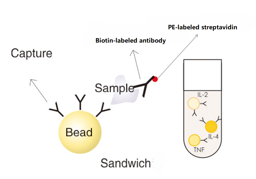
Principle:
Specific antibodies are coupled to specific microsphere surfaces with different fluorescence intensities to prepare capture microspheres (Capture Antibodies) that become the factors to be tested. The capture microspheres are incubated with the sample to be tested (serum, plasma or cell culture medium), at which time the cytokine to be tested is captured by the specific microspheres. The biotin-labeled detection antibody forms an immune complex of antibody-coated microsphere-cytokine-detection antibody, adds phycoerythrin-labeled streptavidin and biotin to combine, detects by flow cytometer to obtain fluorescence intensity of the test substance, the fluorescence intensity is proportional to the content of factors in the sample, and finally combines the standard curves of the twelve cytokine standards to realize quantitative detection of multiple factors in the same sample and assist in judging the immune function state of the body.
Detection sensitivity of different indicators:
|
Specificity |
Detection range |
|
mIL-12P70 |
4.88 pg/mL-5000pg/mL |
|
mTNF-α |
19.53 pg/mL-5000pg/mL |
|
mIFN-r |
19.53 pg/mL-5000pg/mL |
|
mMCP-1 |
19.53 pg/mL-5000pg/mL |
|
mIL-10 |
4.88 pg/mL-5000pg/mL |
|
mIL-6 |
4.88 pg/mL-5000pg/mL |
|
mGM-CSF |
4.88 pg/mL-5000pg/mL |
|
mIL-17A |
4.88 pg/mL-5000pg/mL |
|
mIL-5 |
4.88 pg/mL-5000pg/mL |
|
mIL-1β |
4.88 pg/mL-5000pg/mL |
|
mIL-4 |
4.88 pg/mL-5000pg/mL |
Mouse cytokine detection sensitivity
|
Specificity |
Detection range |
|
human TNF-α |
2.44 pg/mL-5000pg/mL |
|
human IFN-γ |
19.53 pg/mL-5000pg/mL |
|
human IL-2 |
2.44 pg/mL-5000pg/mL |
|
human IL-4 |
1.22 pg/mL-5000pg/mL |
|
human IL-6 |
19.53 pg/mL-5000pg/mL |
|
human IL-5 |
1.22 pg/mL-5000pg/mL |
|
human IL-10 |
1.22 pg/mL-5000pg/mL |
|
human IL-12P70 |
4.88 pg/mL-5000pg/mL |
|
human IL-1β |
9.77 pg/mL-5000pg/mL |
|
human IL-8 |
2.44 pg/mL-5000pg/mL |
|
human IL-17 |
4.88 pg/mL-5000pg/mL |
|
human IFN-α |
2.44 pg/mL-5000pg/mL |
Human cytokine detection sensitivity
Note: If the test result is beyond the test range, use standard/sample/microsphere diluent to dilute the sample by appropriate multiple for re-test; If the test result of the sample to be tested is lower than the lower detection limit or is not detected, it is recommended to directly report it as ≤ minimum test
Template creation method:
1. Microsphere group distribution:
|
Specificity |
Microsphere Location |
Region to which the microspheres belong |
|
human TNF-α |
L1 |
R1 |
|
human IFN-γ |
L2 |
R1 |
|
human IL-2 |
L3 |
R1 |
|
human IL-4 |
L4 |
R1 |
|
human IL-6 |
L5 |
R1 |
|
human IL-10 |
L6 |
R2 |
|
human IL-12P70 |
L7 |
R2 |
|
human IL-1β |
L8 |
R2 |
|
human IL-8 |
L9 |
R2 |
|
human IL-17 |
L10 |
R2 |
|
human IFN-α |
L11 |
R3 |
|
human IL-5 |
L12 |
R3 |
Note: The interior of the microspheres is stained with fluorescent dyes of different intensities.
2. Template establishment: For template establishment, please refer to:
(1) Establish a density map template with X-axis as FSC-H and Y-axis as FSC-A, set the gate, excluding fragments and adherent microspheres, as shown in Figure 1 and P6 below;
(2) Establish a linear density map template with X axis as FSC-H and Y axis as SSC-H, and set the positions R1, R2 and R3 of the mixed microspheres, as shown in Figure 2 below;(3) Two logarithmic density plot templates of PE (X-axis) and APC (Y-axis) are suggested to show the microspheres in R1, R2 and R3, respectively, so that the microspheres can be clearly and obviously distributed on the scatter plot and show the PE fluorescence intensity of each factor. As shown in Figure 3, Figure 4, Figure 5.

3. Analysis of sample test results
Each standard and sample are tested on the computer sequentially, and at least 100 microspheres should be obtained for each microsphere group. Use the standard gradient as the standard curve to calculate the sample test results.
CBA application fields:
2. Tumor research and auxiliary diagnosis, curative effect and prognosis monitoring of tumor diseases;
3. Monitoring of immune diseases such as systemic lupus erythematosus (SLE) and neonatal sepsis;
4. Stem cell research and stem cell therapy monitoring;
5. Immunological research and auxiliary diagnosis and monitoring of immune diseases;
6. Research and auxiliary diagnosis and curative effect evaluation of infectious diseases;
7. Immune function monitoring;
8. Drug efficacy evaluation, drug research and development, vaccine research.
Compared with traditional protein quantification, CBA has the following advantages:
1. Micro detection: One sample can detect multiple factors at the same time, only 25ul.
2. The data is more reliable: multiple indicators come from the same sample
3. Fast detection speed: simultaneous detection of multiple factors, no overnight incubation is required.
4. High sensitivity: as low as 1.22 pg/mL.
Absin CBA Box Advantages:
1. Good performance: Using Biotin-SAV signal amplification system, the sensitivity is higher.
2. Magnetic microspheres: Equipped with magnetic marker plates, easy to operate.
3. Wide application: compatible with a variety of flow cytometers.
4. Easy analysis: FCAP v3.0 analysis software is provided for free.
5. Flexible indicators: Detection indicators can be flexibly combined.
Freely combinable multi-factor detection Kit:
Absin Existing Kit
|
Item number |
Product Name |
Specifications |
|
abs50187 |
Mouse eleven cytokine detection kit (flow fluorescence method) |
48T/96 T |
|
abs50186 |
Human twelve cytokine detection kit (flow fluorescence luminescence method) |
48T/96 T |
Supporting consumables
|
Item number |
Product Name |
Specifications |
|
abs7985 |
96-well magnetic plate |
1 |
On the basis of the existing indicators of the kit, individual indicators can be selected and combined to form a box with fewer indicators (such as four or five cytokines), but human and mouse indicators are not universal. You can contact the business staff to inquire about the price and delivery date of the portfolio Kit.
|
Human indicators |
Mouse Indicator |
|
human TNF-α |
mouse TNF-α |
|
human IFN-γ |
mouse IFN-r |
|
human IL-4 |
mouse IL-4 |
|
human IL-6 |
mouse IL-6 |
|
human IL-5 |
mouse IL-5 |
|
human IL-10 |
mouse IL-10 |
|
human IL-12P70 |
mouse IL-12P70 |
|
human IL-1β |
mouse IL-1β |
|
human IL-17 |
mouse IL-17A |
|
human IL-2 |
mouse MCP-1 |
|
human IL-8 |
mouse GM-CSF |
|
human IFN-α |
|
Absin provides antibodies, proteins, ELISA kits, cell culture, detection kits, and other research reagents. If you have any product needs, please contact us.
|
Absin Bioscience Inc. Email: worldwide@absin.cn |
 Follow us on Facebook: Absin Bio Follow us on Facebook: Absin Bio |