- Cart 0
- English
Marker significance of CD44 and CD62L on tumor cells and T cells
July 01, 2025
Clicks:2786
CD44 and CD62L are two types of molecular markers on the surface of T cells, which play a key role in the regulation of T cell activation, migration and function. Today, Xiaoai will take you to see the labeling effect of these two indicators on tumor cells and T cell species.
CD44 is a transmembrane glycoprotein that belongs to the integrin family. It is widely expressed in a variety of cell types, including leukocytes, endothelial cells, fibroblasts, and certain tumor cells.
Cell adhesion and migration: CD44 is involved in cell adhesion, migration, and signaling by binding to hyaluronic acid (HA). In immune cells, CD44 contributes to the localization and migration of T cells and B cells in lymphoid tissues.
Tumor biology: CD44 is one of the markers of cancer stem cells and is involved in the proliferation, migration and invasion of tumor cells. Tumor cells that express CD44 are generally more metastatic and resistant to drugs.
Immunomodulation: CD44 also plays a regulatory role in the immune response, affecting the activation and function of T cells.
1. Relationship between CD44 and T cells and B cells
Migration and localization: CD44 aids in the migration and localization of T and B cells in lymphoid tissue by binding to hyaluronic acid (HA). In lymph nodes, CD44-mediated cell-matrix interactions contribute to the homing and residency of immune cells.
Immune activation: CD44 also plays a supporting role in the activation of T cells and B cells. By binding to HA, CD44 can enhance the interaction of immune cells with antigen-presenting cells (APCs) and promote the initiation of immune responses.
Adhesion and migration: CD44 regulates the migration and localization of T cells in lymphoid tissues, sites of inflammation, and target tissues through the interaction of adhesion and matrix molecules.
Activation signaling: CD44 interacts with other surface receptors, such as TCRs and costimulatory molecules, to enhance T cell activation signaling and promote cell proliferation and function.
Stored memory: CD44 plays an important role in the formation and maintenance of long-term immune memory, helping T cells to quickly initiate a response when they are again exposed to the same antigen.
Tumor cell markers: CD44 is one of the most common cancer stem cell markers and is involved in the metastasis, adhesion, and migration of tumor cells.
2. Relationship between CD44 and natural killer cells (NK cells).
Cytotoxicity: CD44 also plays a role in the cytotoxicity of NK cells. NK cells with high CD44 expression are more cytotoxic and are able to kill target cells more efficiently.
Tumor microenvironment: In the tumor microenvironment, tumor cells with high CD44 expression can interact with immune cells and affect the function of immune cells. For example, CD44 can inhibit the activity of T cells by binding to HA to form an immunosuppressive microenvironment.
Immune escape: Tumor cells with high CD44 expression have stronger immune escape ability and are able to avoid being recognized and killed by immune cells through a variety of mechanisms.
CD62L (L-selectin) is an adhesion molecule that belongs to the selectin family. It is mainly expressed on the surface of white blood cells, including T cells, B cells, and natural killer cells.
Cell homing: CD62L plays a key role in the homing process of white blood cells, helping them move from the blood to the lymphoid tissue. In the inflammatory response, CD62L mediates the initial adhesion of leukocytes to endothelial cells.
Immune cell localization: CD62L contributes to the localization of T cells and B cells in lymph nodes and is involved in the initiation and regulation of immune responses.
Immune memory: CD62L-high expression T cells often have the properties of central memory T cells (Tcms), which are able to respond quickly when re-encountered with antigens.
3. Relationship between CD62L and T cells and B cells
Homing: CD62L plays a key role in the homing process of white blood cells, helping T cells and B cells to travel from the bloodstream into lymphoid tissue. In the inflammatory response, CD62L mediates the initial adhesion of leukocytes to endothelial cells, promoting the rolling and migration of leukocytes.
Immune response: CD62L contributes to the initiation and regulation of the immune response by promoting the localization of T cells and B cells in the lymph nodes. T cells with high CD62L expression often have the properties of central memory T cells (Tcms), which are able to respond quickly when re-encountered with antigens.
Inactive state recognition: In the inactive state, the expression of CD62L enables T cells to recognize and enter secondary lymphoid organs such as lymph nodes to participate in the initiation of immune responses.
Central memory cell markers: CD62L is considered a marker of central memory T cells, which have high CD62L expression and have the ability to quickly locate to sites such as lymph nodes for rapid reresponse.
4. Relationship between CD62L and natural killer cells (NK cells).
Migration and localization: CD62L is also involved in the migration and localization of NK cells, helping NK cells to aggregate at the site of inflammation and enhancing their cytotoxic functions.
Immune cell distribution: CD62L affects the strength and duration of immune responses by regulating the distribution of immune cells in different tissues. For example, T cells with high CD62L expression reside longer in the lymph nodes and are more effectively involved in the immune response.
5. Distinguish T cell subsets by CD44 and CD62L
Naïve T cells: CD44low/- CD62L+
Central memory T cells (Tcm): CD44high CD62L+
Effector memory T cells (Tem) or effector T cells (Teff): CD44high CD62L-
6. Additional markers are used to distinguish Tem from Teff
CD127 (IL-7Rα): Tem is normally expressed, while Teff expression is reduced.
KLRG1: Teff was highly expressed, Tem expression was low.
CD69: Teff hyperexpression in the early stages of activation.
Bcl-2:Tem expression was higher and Teff expression was lower.
Cytokine production: Detection of IFN-γ, TNF-α, IL-2, and more.
Proliferative capacity: Tem generally has a stronger proliferative potential.
With these markers and functional assays, different types of T cells can be more accurately distinguished, leading to a better understanding of immune responses and disease mechanisms.
7. Potential applications of CD44 and CD62L in the treatment of tumors
Drug delivery: CD44 is the receptor for hyaluronic acid (HA), which can act as a drug carrier to deliver cytotoxic drugs or prodrug convertases around tumor cells, forming a cytotoxic fence that enhances anti-tumor effects. For example, HA-modified liposomes exhibit excellent antitumor activity in vitro and in murine tumor models.
Antibody conjugates: Anti-CD44 antibodies can be used to deliver radioisotopes or cytotoxic drugs to treat CD44-expressing tumors. In early clinical trials, patients with breast or head and neck tumors treated with anti-CD44 conjugates were stable.
Enhance immune response: Tumor cells with high CD44 expression can be treated with combination of immune checkpoint inhibitors (such as PD-1/PD-L1 inhibitors) to enhance the anti-tumor effect. For example, the MUSIC treatment strategy combined with PD-1 inhibitors not only showed stronger primary tumor suppression, but also greatly slowed tumor metastasis, with a 76% increase in median survival time in mice in the combination treatment group.
Predicting immunotherapy response: CD44 can be used as a promising biomarker for predicting immunotherapy response and mediating PD-L1 expression. In bladder cancer, high CD44 expression was associated with poor prognosis and PD-L1 expression.
Enhance T cell infiltration: CD62L-positive T cells can enter secondary lymphoid organs such as lymph nodes and participate in the immune response. By regulating the expression of CD62L, the infiltration of T cells in the tumor microenvironment can be enhanced, thereby improving the efficacy of immunotherapy.
Combination therapy: T cells with high expression of CD62L exhibited stronger anti-tumor effects in combination therapy. For example, the MUSIC treatment strategy combined with PD-1 inhibitors not only enhanced the inhibition of the primary tumor, but also significantly slowed tumor metastasis.
Modulates immune cell distribution: CD62L can regulate the distribution of T cells in the tumor microenvironment, and by increasing the number of CD62L-positive T cells, immune surveillance and anti-tumor responses can be enhanced.
summary
CD44 is mainly involved in cell adhesion, migration and tumor biology, and is a marker of cancer stem cells, which has important clinical application value. Due to its high expression in cancer stem cells, CD44 has emerged as a potential target for tumor therapy. By targeting CD44, it can inhibit the proliferation and metastasis of tumor cells and enhance the anti-tumor effect. In tumor therapy, it mainly plays a role in targeted drug delivery, enhancing immune responses, and modulating the tumor microenvironment. Its high expression is associated with the poor prognosis of a variety of tumors, and is expected to become an important target for tumor treatment.
CD62L is mainly involved in the homing of leukocytes and the localization of immune cells, and is a marker of central memory T cells, which is of great significance for immunotherapy. In tumor treatment, the effect of immunotherapy is mainly improved by enhancing T cell infiltration and regulating the distribution of immune cells. In CAR-T cell therapy, the efficacy and durability of CAR-T cells can be improved by selecting T cells with high CD62L expression. Through combination therapy, the anti-tumor effect can be significantly enhanced and tumor metastasis can be slowed.
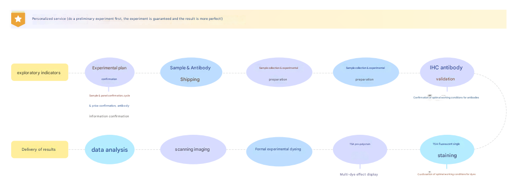
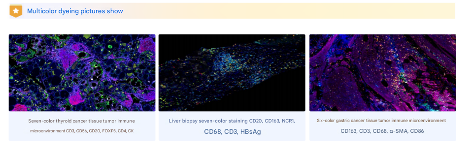
Abics mIHC Multicolor Assay Service
Multiplex fluorescence immunohistochemistry services with up to 9 indicators and 10 colors (including DAPI), with sample species covering human, mouse, rat, pig, etc.; Tumor samples are the majority of tissue types, covering major organs such as lung, stomach, liver, kidney, intestine, thyroid, pancreas, labial gland, lacrimal gland, etc., as well as special samples such as eyeballs, nerves, and brains. Slide types cover paraffin sections, tissue chips (TMA), frozen sections, large tissue sections, thick sections, etc., and provide full-slide scanning and quantitative analysis.
Absin provides antibodies, proteins, ELISA kits, cell culture, detection kits, and other research reagents. If you have any product needs, please contact us.
|
Absin Bioscience Inc. Email: worldwide@absin.net |
 Follow us on Facebook: Absin Bio Follow us on Facebook: Absin Bio |