- Cart 0
- English
Characteristics of 'Black Jelly Worm' Contamination in Cell Culture II
January 10, 2025
Clicks:2396
Last time in Exploring Black Glue Worms (Part 1), we mainly discussed the situation where some black glue worm samples were identified as bacteria. Additionally, some samples were identified as mycoplasmas.Here are two typical identification results of black glue worms to share with everyone:
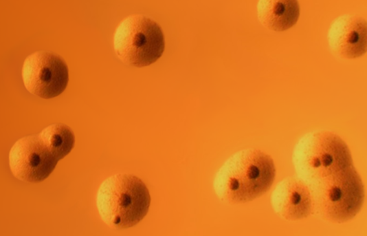
Figure 1 Colonies of Mycoplasma hyorhinis on solid culture medium
Identification Results: Samples 7, 12, and 18 were identified as Mycoplasma hyorhinis (Swine Mycoplasma)
Size: 0.15-0.3um
Host of Infection: It infects the epithelial cells of the upper respiratory tract of pigs, transmitted through the oral and nasal routes. Piglets can be infected by sows or their peers (asymptomatic carriers).
Route of Cell Contamination: It is speculated that trypsin is mainly extracted from pig pancreas, purified to obtain crystals, and then made into a lyophilized preparation. Improperly filtered trypsin solution may carry swine mycoplasma, so it is recommended to purchase trypsin from well-known brands to reduce the risk of contamination.
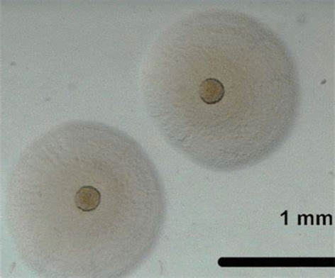
Figure 2 Colonies of Mycoplasma bovis on solid culture medium
Identification Results: Sample 23 was identified as Mycoplasma bovis (Bovine Mycoplasma);
Size: 0.15-0.3um
Host of Infection: Mainly parasitizes within cattle, with a wide range of infection sites, including the respiratory tract, causing pneumonia, otitis media, arthritis, and conjunctivitis.
Route of Cell Contamination: One of the main nutrients for cell proliferation is fetal bovine serum. Fetal bovine serum is derived from animals and is primarily filtered and sterilized through a 0.1um filter membrane. If the blood source and filtration are not properly done, it can potentially introduce mycoplasma contamination to the cells. Therefore, it is recommended to purchase fetal bovine serum from well-known brands.
Next, let's analyze the common characteristics of black glue worm contamination that we introduced in the last episode: active swimming, rotating movement, and trembling in place, these three are the most common contamination phenomena. Mycoplasmas, unlike bacteria, do not have flagella for movement, obviously it is not likely to be active swimming or rotating movement. Samples identified as mycoplasma contamination are very likely to be this type of black glue worm sample - the black glue worms we see trembling in place in the liquid. Moreover, mycoplasmas are currently known as the smallest prokaryotic organisms, which can only be observed under an electron microscope, the trembling black dots we see are very likely to be mycoplasma colonies.
First, let's look at several videos of "black glue worms" that might be mistaken for "trembling in place" by everyone:
Video 1 Trembling black dots (Video of L02 liver cell line contaminated with black glue worms)
Video 2 Trembling black dots
Video 3 Trembling black dots
Video 4 Trembling black dots
From these "black glue worm" contamination videos, we can summarize the morphological characteristics of black glue worms:
Almost all are black and oval-shaped dots, varying in size; they float outside the cells, trembling, and upon close observation, one can see that they are trembling in place.
Furthermore, in our analysis of cell contamination for clients, we have also found that the so-called "black glue worm" contamination is really "another world". Below is a screenshot of our chat analysis:
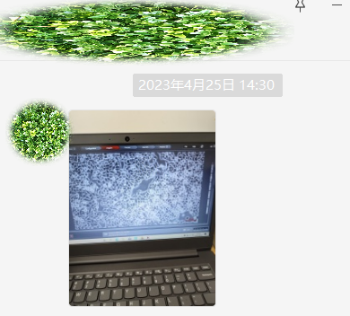
Figure 3 Cell image from client's chat screenshot
Zooming in on this image as follows:
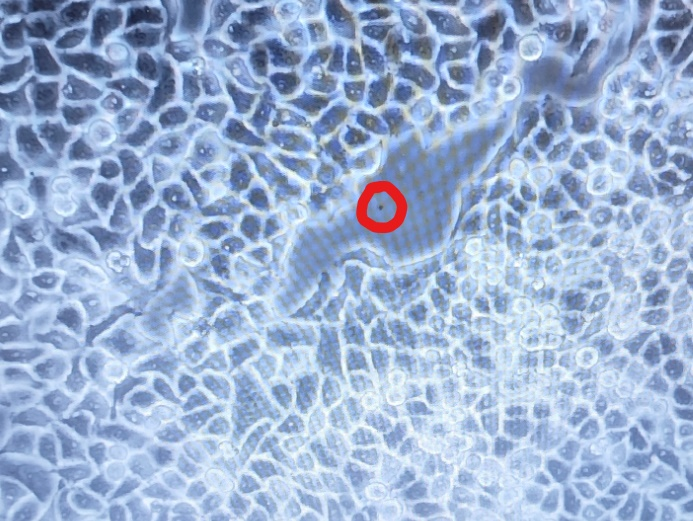
Figure 4 The black dots within the red circle are very likely to be mycoplasma colonies
Next, to further confirm, we asked the client to send a video:
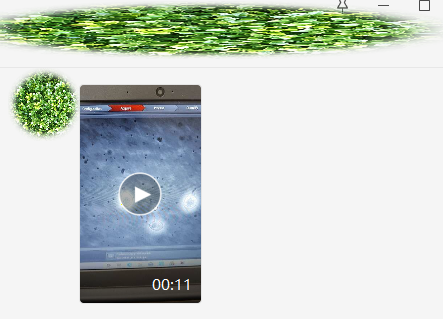
Figure 5 Cell video from client's chat screenshot
Let's zoom in on the video for observation:
Video 6 Trembling black dots
From the video, we can see that many oval-shaped black dots are trembling in place. Previously, in our cell communication group, we asked the client whether they had tested for mycoplasmas, the client said they had, and it was confirmed to be mycoplasma contamination.
Through this case analysis, we speculate that the commonly referred to "trembling" "black glue worm" contamination is very likely to be mycoplasma contamination, and these trembling small round dots are very likely to be mycoplasma colonies. In the future, we will collect a large number of these trembling black dot samples, conduct sequencing identification, and further confirm our speculation with big data, sharing these interesting results with everyone.
If your cells have indeed been "hit", do not fear, our mycoplasma elimination reagent will clear the way and protect your cells.
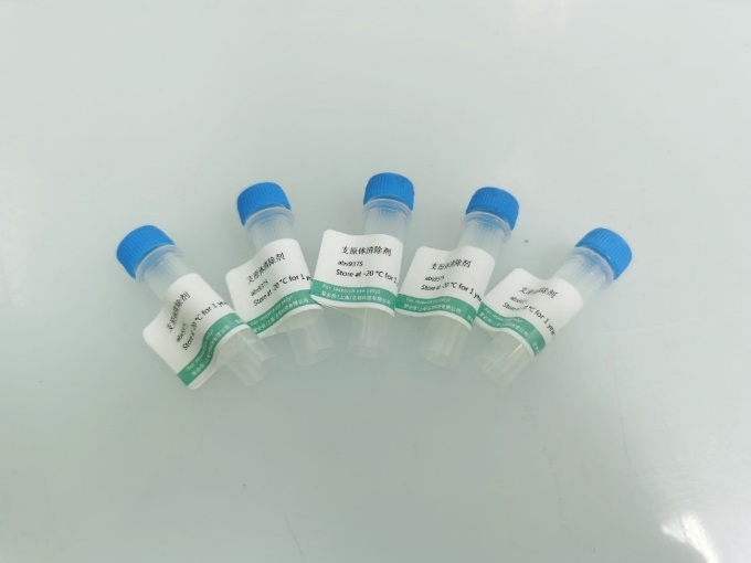 Mycoplasma Removal Reagent abs9375-200uL×5 |
Product Advantages: 1. Specifically eliminates mycoplasmas from the culture medium; 2. Effectively removes mycoplasmas in just 3-6 days; 3. Peptide-based active ingredients do not lead to resistance; 4. Virtually non-toxic to cells, tested on over 100 types of cells, including but not limited to HEK293, HeLa, MCF-7, MRC-5, NIH-3T3, CCC-ESF, CHO-S, CHO-K1, CHO-DG44, H295R, HL-60, K562, MDA-MB-231, SP2/0, T47D, BM, and BV2, etc.; 5. Broad-spectrum, can replace double antibiotics, capable of clearing common Gram-negative and Gram-positive bacteria; 6. Can be directly added to serum or culture medium for mycoplasma contamination removal. |
This concludes our discussion for this session. If you have any cell culture issues, feel free to scan the code to join our WeChat group for communication.
|
Absin Bioscience Inc. |
 Follow us on Facebook: Absin Bio Follow us on Facebook: Absin Bio |