- Cart 0
- English
Ferroptosis Mechanism and Detection Methods
December 24, 2024
Clicks:521
What is ferroptosis?
Ferroptosis is a form of oxidative cell death induced by small molecules, characterized by its dependence on iron ions. Research has shown that ferroptosis is associated with a variety of diseases, including neurodegenerative diseases, tumors, atherosclerosis, diabetes, and acute kidney injury. The pathogenic mechanisms are complex, and diseases can be intervened by activating or inhibiting ferroptosis. Therefore, ferroptosis has become a hot topic of research in recent years.
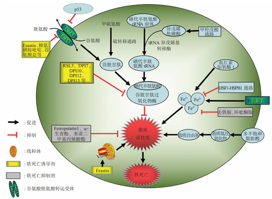
Figure 1: Mechanism of Ferroptosis[1]
Ferroptosis Mechanism
Ferroptosis is caused by the accumulation of lipid peroxides dependent on ferrous ions and reactive oxygen species (ROS). The mechanism of ferroptosis is depicted in Figure 1. The upstream pathway involves directly or indirectly affecting the activity of glutathione peroxidases (GPXs), which reduces the cell's antioxidant capacity, leading to increased lipid peroxidation, more lipid ROS, and the initiation of ferroptosis. Additionally, under the influence of ferrous ions or lipoxygenases, highly expressed unsaturated fatty acids on the cell membrane undergo lipid peroxidation, inducing cell death. The essence of ferroptosis is the depletion of glutathione and the decline in the activity of glutathione peroxidase 4 (GPX4). Lipid peroxides cannot be metabolized through the glutathione reductase reaction catalyzed by GPX4, after which ferrous ions oxidize lipids to produce reactive oxygen species, thereby promoting the occurrence of ferroptosis.
Characteristics of Ferroptosis Morphology
Currently, the more extensively studied modes of cell death include ferroptosis, apoptosis, necrosis, and autophagy, which can be distinguished morphologically as shown in Figure 2.
Ferroptosis: Mitochondria shrinkage, increased membrane density, and reduction or disappearance of mitochondrial cristae;
Apoptosis: Chromatin condensation, nuclear pyknosis, and fragmentation; formation of apoptotic bodies, with no significant changes in mitochondrial morphology;
Necrosis: Swelling of organelles, damage to the plasma membrane, cell lysis, leakage of cellular contents, and tissue damage;
Autophagy: Formation of double-membrane autophagosomes (containing cytoplasmic components such as mitochondria, endoplasmic reticulum, ribosomes, etc.) or single-membrane autolysosomes (where cytoplasmic components have been degraded).
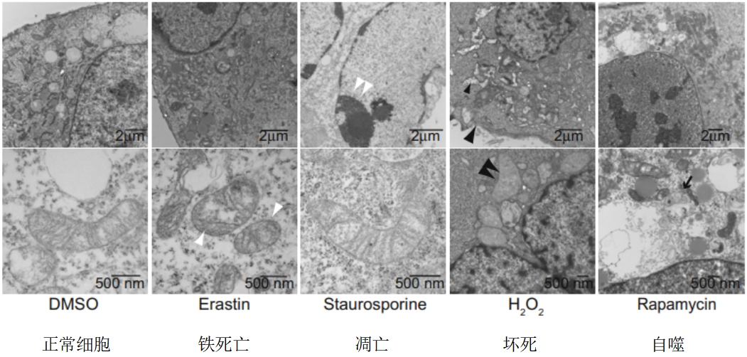
Figure 2: Morphological Symptoms of Cellular Ferroptosis, Apoptosis, Necrosis, and Autophagy[2]
Regulatory Pathways of Ferroptosis
Studies have shown that ferroptosis can be induced by a variety of compounds (Table 1), and its occurrence is regulated by pathways such as the cystine-glutamate antiporter (System xc-), p53, GPX4, and voltage-dependent anion channels (VDACs). In addition, the induction of lipid peroxidation reactions can also lead to ferroptosis; heme oxygenase-1 (HO-1) and transferrin, which are sources of intracellular iron, are also involved in the regulation of ferroptosis[3]。
Table 1: Ferroptosis Inducers and Inhibitors
|
Ferroptosis Inducers |
Ferroptosis Inhibitors |
||
|
Compounds |
Mechanisms |
Compounds |
Mechanisms |
|
Erastin |
System xc-,VDAC2/3 |
Trolox |
Lipophilic Antioxidants |
|
RSL3 |
GPX4 |
Glutathione |
Oxidative Pathway/Intracellular Iron |
|
Sulfasalazine |
System xc- |
Ferrostatin-1 |
ROS, Lipid Peroxidation |
|
Sorafenib |
System xc- |
Liproxstatin-1 |
ROS, Lipid Peroxidation |
|
Artesunate |
氧化途径 |
Deferoxamine Mesylate |
Iron Chelator |
|
Cisplatin |
GSH |
Deferasirox |
Iron Chelator |
|
FIN56 |
GPX4 |
Zileuton |
Lipoxygenase Inhibitor |
|
BAY 87-2243 |
ROS |
PD-146176 |
Lipoxygenase Inhibitor |
Diagnosis and Detection of Ferroptosis
When cells undergo ferroptosis, they exhibit biochemical characteristics such as iron accumulation, lipid peroxidation, and a decrease in mitochondrial membrane potential. Therefore, ferroptosis can be diagnosed and detected by measuring specific elements, including:
· Glutathione peroxidase 4 (GPX4) and glutathione (GSH)· Reactive oxygen species (ROS) and lipid peroxidation
· Iron ion levels
· Mitochondrial activity
These multiple approaches can be used to comprehensively verify the occurrence of ferroptosis [4].
1. Methods for Detecting GPX4 and GSH:
(1) Spectrophotometric assays to evaluate GPX4 activity;
(2) Detection using an anti-GPX4 antibody.
2. Methods for Detecting ROS and Lipid Peroxidation:
(1) Semi-quantitative analysis with specific fluorescent probes (such as DCHF-DA);
(2) ELISA and TBAR assays for detecting downstream byproducts of lipid ROS (such as 4-HNE protein adducts and MDA);
(3) Inhibitors: Use of specific antioxidant ferroptosis inhibitors, such as Ferrostatin-1, for correlative analysis.
Iron Ion Levels:
(1) The level of iron ions can be detected using colorimetric reactions with compounds such as phenol red, which changes color upon binding with iron;
(2) Chelating agents or genetic manipulations that affect iron ion concentration (such as transferrin receptor, ferritin, and mitochondrial ferritin) can also influence ferroptosis.
Mitochondrial Detection:
(1) Observation of characteristic morphological changes in mitochondria during ferroptosis, including increased membrane density, reduced cristae, and decreased membrane potential;
(2) After a decrease in GPX4 function, cellular fragmentation may occur within a few hours. The morphology of mitochondria can be detected using mitochondrial markers such as MitoTracker Green FM or similar dyes. (Note: MitoScene Green I seems to be a typo or a less commonly used dye; a more standard dye like MitoTracker Green FM is suggested for clarity.)
Ferroptosis, as a novel form of programmed cell death, has increasingly been recognized for its significant role in disease research in recent years. Although a preliminary understanding of its characteristics and mechanisms has been established, many questions still remain to be addressed. These include further elucidating the detailed regulatory mechanisms of ferroptosis in various diseases and developing new technologies for detecting specific biomarkers of ferroptosis.
References
[1] 周文博,孔晨飞,秦高伟,等.铁死亡发生机制的研究进展[J].生物化学与生物物理进展, 2018, 45(1):7.
[2] Dixon SJ, Lemberg KM, Lamprecht MR, et al. Ferroptosis: an iron-dependent form of nonapoptotic cell death[J]. Cell, 2012, 149( 5): 1060-1072.
[3] Gao M, Monian P, Quadri N, et al. Glutaminolysis and transferrin regulate ferroptosis. Mol Cell, 2015, 59(2): 298-308.
[4] 孙悦,付松波,李亦兰.心肌细胞铁死亡及其检测方法[J].心血管病学进展, 2023, 44(2):167-171.
Ferroptosis Research Detection Kits:
|
Item NO. |
Product Name |
Size |
|
Malondialdehyde Microplate Assay Kit |
96T |
|
|
Iron Microplate Assay Kit |
96T |
|
|
TBARS Microplate Assay Kit |
96T |
|
|
Glutathione Microplate Assay Kit |
96T |
|
|
Glutathione Reductase Microplate Assay Kit |
96T |
|
|
Glutathione Peroxidase Microplate Assay Kit |
96T |
Absin provides antibodies, proteins, ELISA kits, cell culture, detection kits, and other research reagents. If you have any product needs, please contact us.
|
Absin Bioscience Inc. |
 Follow us on Facebook: Absin Bio Follow us on Facebook: Absin Bio |