- Cart 0
- English
mIHC Literature Interpretation: Exploring the Tumor Immune Microenvironment of Melanoma
November 26, 2024
Clicks:625
Melanoma:
A tumor that arises from the transformation of melanocytes, which are normal pigment-producing cells found in the basal layer of the skin's epidermis. These cells produce melanin and transfer it to surrounding keratinocytes, where the pigment resides in the nuclei of these cells to protect the chromosomes from damage caused by exposure to light radiation. When exposed to long-term ultraviolet (UV) radiation from sunlight, individuals with fair skin and poor UV protection may experience genetic mutations in their normal melanocytes, leading to the development of malignant cells that can reproduce and expand indefinitely, resulting in melanoma. Cutaneous melanoma (CM) is more common in Caucasians, while the incidence in Asians and African descent populations is relatively lower. However, melanoma is not limited to this form. Studies have shown that in Asian populations, acral melanoma (AM), which occurs on the fingers, toes, and soles of the feet, accounts for approximately 50% of melanoma cases in East Asia. Since these areas have minimal exposure to UV light, the etiology of AM differs from that of CM. The distinct pathogenesis also leads to the fact that some immunotherapies effective for CM do not perform as well in patients with AM.
To better understand acral melanoma, on November 25, 2022, Yang Jilong, Li Xiangchun, and Chen Kexin from Tianjin Medical University co-corresponded in Nature Communications to publish a research paper titled "A single-cell analysis reveals tumor heterogeneity and immune environment of acral melanoma". They conducted single-cell RNA sequencing on 63,394 cells from 5 acral melanoma (AM) and 3 cutaneous melanoma (CM) samples to study the heterogeneity of the tumor and the immune environment. The study found that compared to CM, AM has a distinct immunosuppressive state, with high expression of PD-1 and TIM-3 in the exhausted CD8+ T cells of AM.
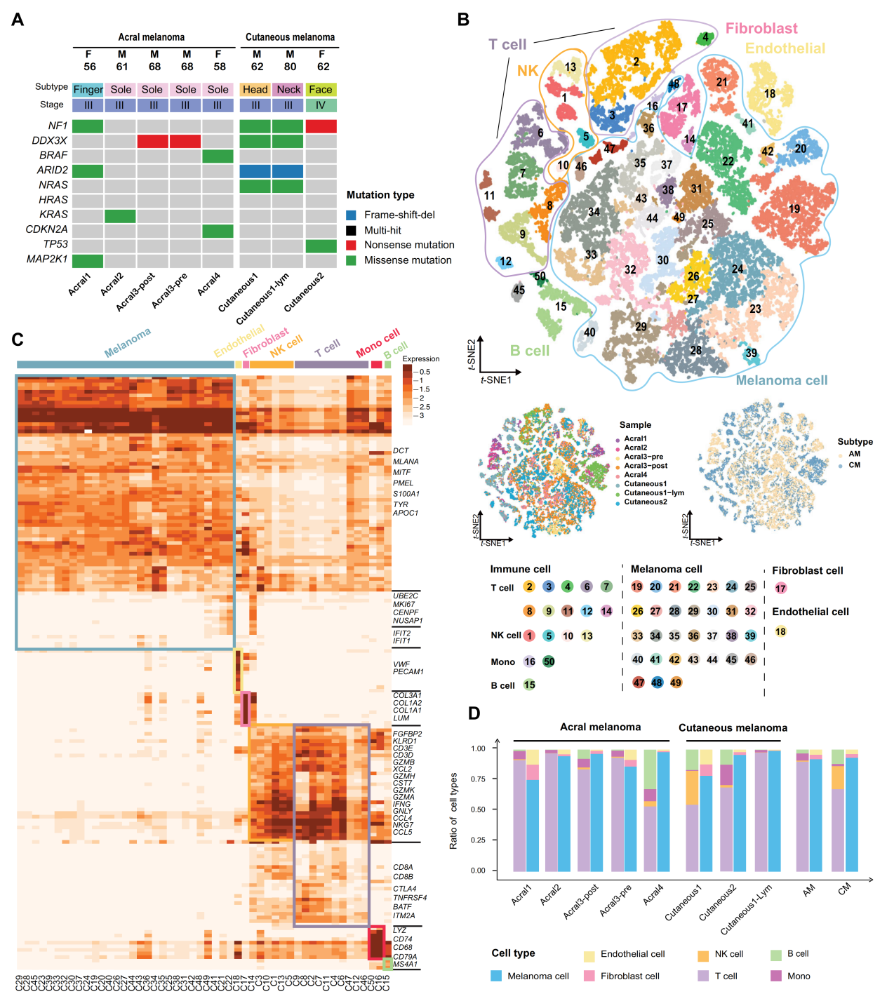
Single-cell RNA sequencing analysis of the tumor microenvironment in AM and CM
In addition to single-cell sequencing, the research team also used the Absin Pentachrome Multiplex Fluorescent Immunohistochemistry Staining Kit (abs50013) for multiplex staining of relevant immune targets in situ on tissue sections, further substantiating their findings at the protein level. The results showed that CD8+ T cells in acral melanoma (AM) that are in an exhausted state co-express high levels of TIM-3 and PD-1 (Figure 1); after monotherapy with anti-PD-1 immunotherapy, the proportion of CD8+ T cells in AM patients decreased compared to before treatment, while the proportion of CD4+ T cells and B cells increased, indicating that monotherapy with anti-PD-1 is ineffective for AM patients (Figure 2).
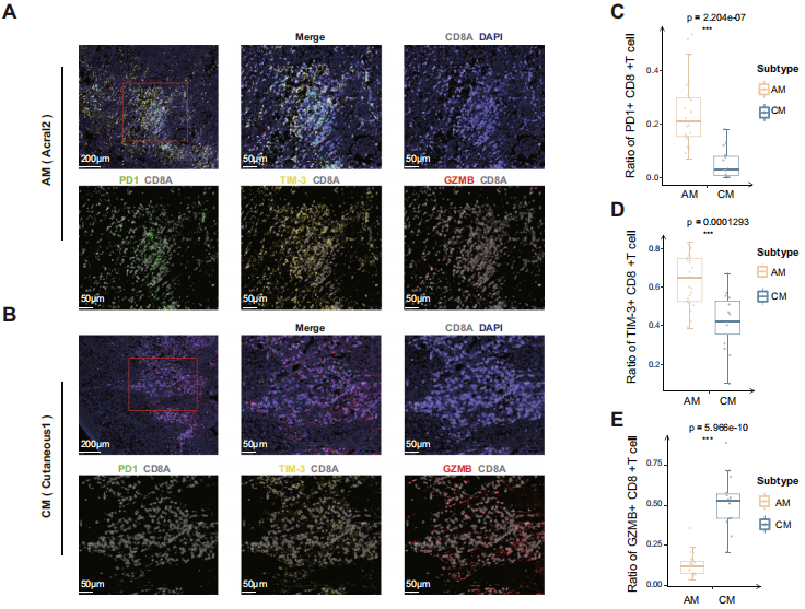
Figure 2
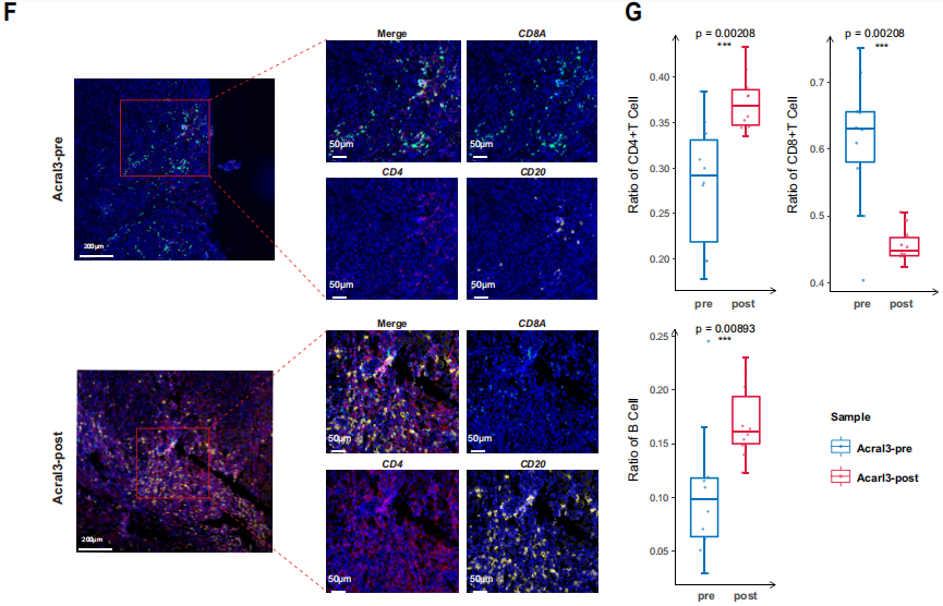
Figure 3
In addition to exploring the immune microenvironment of AM, the research team also found that the combined application of anti-PD-1 and anti-TIM-3 drugs significantly increased the apoptosis ratio of AM tumor cells; immunotherapy combined with genes related to the EGFR pathway may also improve the efficacy of immunotherapy in AM patients. This provides valuable information for the development of more effective therapeutic targets and immune therapy-related biomarkers for acral melanoma patients.
References
Zhang C, Shen H, Yang T, Li T, Liu X, Wang J, Liao Z, Wei J, Lu J, Liu H, Xiang L, Yang Y, Yang M, Wang D, Li Y, Xing R, Teng S, Zhao J, Yang Y, Zhao G, Chen K, Li X, Yang J. A single-cell analysis reveals tumor heterogeneity and immune environment of acral melanoma. Nat Commun. 2022 Nov 25;13(1):7250. doi: 10.1038/s41467-022-34877-3. PMID: 36433984; PMCID: PMC9700682.
|
Item NO. |
Product Name |
Size |
|
Absin 4-Color IHC Kit (Anti-Rabbit and Mouse Secondary Antibody) |
20T/100T |
|
|
Absin 4-Color IHC Kit(Anti-Rabbit Secondary Antibody) |
20T/100T |
|
|
Absin 5-Color IHC Kit (Anti-Rabbit and Mouse Secondary Antibody) |
20T/100T |
|
|
Absin 5-Color IHC Kit (Anti-Rabbit Secondary Antibody) |
20T/100T |
|
|
Absin 6-Color IHC Kit (Anti-Rabbit and Mouse Secondary Antibody) |
20T/100T |
|
|
Absin 6-Color IHC Kit (Anti-Rabbit Secondary Antibody) |
20T/100T |
|
|
Absin 7-Color IHC Kit (Anti-Rabbit and Mouse Secondary Antibody) |
20T/100T |
|
|
Absin 7-Color IHC Kit(Anti-Rabbit Secondary Antibody) |
20T/100T |
|
|
Antibody eluent (for mIHC) |
30ml |
Absin provides antibodies, proteins, ELISA kits, cell culture, detection kits, and other research reagents. If you have any product needs, please contact us.
|
Absin Bioscience Inc. |
 Follow us on Facebook: Absin Bio Follow us on Facebook: Absin Bio |