- Cart 0
- English
BD single-cell sequencing with Absin multicolor to explore brain tumor microenvironment
November 14, 2024
Clicks:689
Brain malignant tumors, commonly referred to as brain cancers, are divided into two major categories: primary and secondary. Primary brain malignant tumors, with gliomas being the most common, are tumors that originate from brain glial cells. Secondary tumors mainly refer to metastatic brain tumors formed by the spread of cancer from other parts of the body to the brain, with an incidence rate about 10 times that of primary tumors; lung cancer is one of the common primary cancer types.
On November 6, 2022, Professor Jin Wei and Professor Cao Yiqun's team from Fudan University Cancer Hospital published an article titled "Single-cell RNA sequencing reveals cellular and molecular reprogramming landscape of gliomas and lung cancer brain metastases" in Clin. Transl. Med (IF=8.554). Using scRNA-seq technology (BD Rhapsody system) combined with multiplex fluorescence immunohistochemistry (abs50015, Absin), they analyzed the heterogeneity of GM and LC cells and identified a group of stem-like cells with specific molecular markers, which has significant guiding significance for the immunotherapy of lung cancer brain metastasis.

The full process of sampling, sequencing, analysis, and in situ tissue validation.
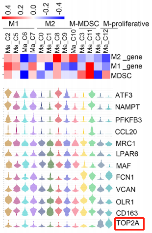
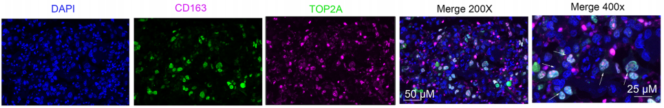
In the study, scientists found that a subset of proliferating macrophage clusters was associated with poor prognosis. The bubble chart illustrates the expression of TAM-related genes in resident microglia of the central nervous system (CNS) and monocyte-derived macrophages (MDMs), with CD163 highly expressed in MDMs and significantly low in CNS. Violin plot analysis revealed that the distribution of the TOP2A gene in proliferating macrophages (last two columns) had better extremes and median values compared to other cells. Subsequent research teams validated the cellular clusters at the tissue protein level by performing multiplex immunofluorescence staining of TOP2A and CD163.
 |
 |
 |
|
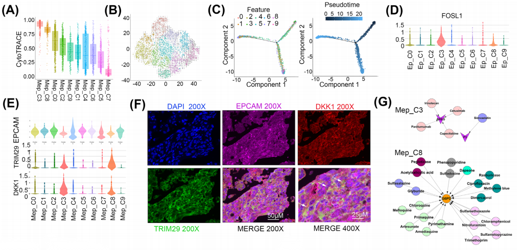
Cancer stem cells (CSCs) are a subset of cells that can lead to cancer metastasis and resistance to chemotherapy or radiotherapy. The research team identified malignant cells through copy number variation analysis and classified them into 10 malignant epithelial cell subpopulations. Based on CytoTRACE analysis and pseudotime analysis, the malignant epithelial cell subpopulations Mep_C3 and C8 showed lower differentiation levels, potentially containing CSCs. To confirm the presence of Mep_C3 and C8 in lung cancer (LC) tissue, the team performed multiplex immunohistochemistry to detect the expression of key markers (EPCAM/DKK1/TRIM29), finding that DKK1 and TRIM29 were specifically enriched in these cells.
Overall, the team's research findings provide particular insights into the characteristics of microglia, macrophages, neutrophils, ECs, and T cells, and also identify a subset of cancer stem cells. This will deepen the understanding of the brain tumor microenvironment, thereby offering some improved approaches for patient management in precision medicine.
Multiplexing refers to multiplex fluorescence immunohistochemistry (mIHC), which is primarily based on the Tyramide Signal Amplification (TSA) technique. TSA is an enzymatic detection method that utilizes horseradish peroxidase (HRP) for high-density in situ labeling of target proteins or nucleic acids. This technique is compatible with high-performance dyes such as Alexa Fluor®, traditional fluorescent dyes, and colorimetric detection systems.
The main principle of TSA involves the peroxidase reaction of tyramide. Tyramide reacts with HRP in the presence of hydrogen peroxide (H2O2) to form covalent binding sites, generating a large amount of enzymatic products that can bind to surrounding protein residues (including tryptophan, histidine, and tyrosine residues). This results in significant biotin deposition at the antigen-antibody binding sites, which subsequently binds to streptavidin-HRP/fluorophore. Through multiple cycles of this amplification process, a large number of enzyme molecules or fluorophores can be attached, resulting in a geometric amplification of the detection signal.
In simple terms, this method for performing multiplex fluorescence staining utilizes HRP attached to secondary antibodies (instead of directly conjugated fluorophores) to catalyze the activation of non-active fluorophores added to the system. The fluorophore is activated by the action of HRP and hydrogen peroxide, covalently coupling with the tyrosine residues of nearby proteins, thus stabilizing the binding of the protein sample with the fluorophore. Microwave treatment is then applied to wash away the non-covalently bound antibodies, leaving the covalently bound fluorophores on the sample. A different primary antibody is then introduced for a second incubation, and this process is repeated. Once all antibody incubations are complete and the fluorophores are bound, the final detection of results can be performed.

TSA Multiplex Fluorescence Immunohistochemistry Staining Schematic Diagram
|
Item NO. |
Product Name |
Size |
|
Absin 4-Color IHC Kit (Anti-Rabbit and Mouse Secondary Antibody) |
20T/100T |
|
|
Absin 4-Color IHC Kit(Anti-Rabbit Secondary Antibody) |
20T/100T |
|
|
Absin 5-Color IHC Kit (Anti-Rabbit and Mouse Secondary Antibody) |
20T/100T |
|
|
Absin 5-Color IHC Kit (Anti-Rabbit Secondary Antibody) |
20T/100T |
|
|
Absin 6-Color IHC Kit (Anti-Rabbit and Mouse Secondary Antibody) |
20T/100T |
|
|
Absin 6-Color IHC Kit (Anti-Rabbit Secondary Antibody) |
20T/100T |
|
|
Absin 7-Color IHC Kit (Anti-Rabbit and Mouse Secondary Antibody) |
20T/100T |
|
|
Absin 7-Color IHC Kit(Anti-Rabbit Secondary Antibody) |
20T/100T |
参考文献:
Sun HF, Li LD, Lao IW, Li X, Xu BJ, Cao YQ, Jin W. Single-cell RNA sequencing reveals cellular and molecular reprograming landscape of gliomas and lung cancer brain metastases. Clin Transl Med. 2022 Nov;12(11):e1101. doi: 10.1002/ctm2.1101. PMID: 36336787; PMCID: PMC9637666.
Absin provides antibodies, proteins, ELISA kits, cell culture, detection kits, and other research reagents. If you have any product needs, please contact us.
|
Absin Bioscience Inc. |
 Follow us on Facebook: Absin Bio Follow us on Facebook: Absin Bio |