- Cart 0
- English
3D Tumor Spheroid & Organoid Drug Sensitivity Activity Detection Guide
November 07, 2024
Clicks:1011
Overview
In recent years, the use of 3D tumor spheroids and organoid models in high-throughput drug screening of large compound libraries, as well as in testing single or multiple drug concentrations, has demonstrated the ability to help researchers quickly screen for drugs with strong drug sensitivity, good response, and superior efficacy in cancer indications. In 2D cell drug sensitivity activity testing, the CCK8 method is widely used. However, in the drug sensitivity activity of 3D tumor spheroids and tumors, the ATP luminescence method is more accurate than the CCK8 method.
Principle of ATP Bioluminescence Technology
This kit uses a bioluminescent method, which utilizes the catalytic conversion of the substrate luciferin by Firefly luciferase (firefly luciferase) to efficiently utilize the energy of ATP and emit photons. The luminescence signal is directly proportional to the amount of ATP present, and ATP is directly proportional to the number of cells in the organoids, effectively detecting the activity of 3D tumor spheroids and organoids.

This kit is designed for multi-well plates and is an ideal choice for automated high-throughput screening (HTS) of 3D tumor spheroids and organoid proliferation and toxicity analysis. The homogeneous detection step involves directly adding a single reagent to the well plate containing the organoid culture medium, without the need to remove the matrix gel.
The "add-reagent-mix-detect" operation scheme of homogeneous detection ensures that the lysis of 3D tumor spheroids and organoids and the resulting luminescence signal are proportional to the amount of ATP present, which in turn is directly proportional to the number of cells in the organoids. The unique homogeneous detection scheme avoids errors that may be introduced by ATP detection methods requiring multiple steps.
Composition
|
Composition |
10mL |
100mL |
Storage |
|
Organoid ATP Dye |
1 vial |
10 vials |
2-8°C, protected from light |
|
Organoid ATP Fluorescent Dye Buffer |
10mL |
10×10mL |
2-8°C, protected from light |
Attention: It is recommended to prepare and use the product immediately.
Product Features
(1) Simplified organoid activity detection steps: The homogeneous "add-reagent-mix-detect" scheme reduces the operational steps required by other similar detections;
(2) Less organoid usage: Accurately detects cell numbers below the detection limits of commonly used colorimetric and fluorescence methods. Reduces the number of cells required per detection reaction;
(3) Rapid results: Data can be obtained within 10 minutes after adding the reagent;
(4) Flexible detection options: Suitable for various types of multi-well plate operations. Data can be recorded using a luminescence detector or a CCD imaging device;
(5) Continuous processing of culture plates: The luminescence signal is stable, allowing for batch processing of samples.
Usage Method
1. Organoid preparation:
Use a 96-well plate suitable for chemiluminescence detection (transparent bottom and lid, opaque well walls, recommended catalog number: abs7242), seed 5uL-10uL of organoid matrix gel suspension per well, and place in the incubator for more than half an hour. After solidification, add 100uL of organoid culture medium (for 384-well plates, seed 2.5μL-4μL of organoid matrix gel suspension per well, the specific amount depends on the type of 384-well plate), and ensure that the number of cells per well does not exceed 50,000 (for 384-well plates, it is advisable to keep it under 10,000). Set up negative control wells without organoids but with matrix gel culture medium, and culture organoids according to standard organoid culture methods. If necessary, treat organoids with drugs. Additionally, if required, set up a concentration gradient of organoids to determine the effectiveness of the kit later.
2. Reagent preparation:
Add one vial of lyophilized powder to one vial of 10mL buffer, mix well, and keep it in the dark until ready to use.
Note: Use as soon as possible after dissolution, store at -80°C in the dark, and the shelf life is one month.
(1) Dissolve the Organoid ATP Luminescence Detection Reagent, and if necessary, aliquot the reagent appropriately;
(2) Take an appropriate amount of Organoid ATP Luminescence Detection Reagent, 100μL per well for a 96-well plate (25μL per well for a 384-well plate), and equilibrate to room temperature.
3. Organoid viability detection:
(1) Take out the organoid culture plate and equilibrate at room temperature for 10 minutes (usually not exceeding 30 minutes);
(2) Remove the organoid culture medium from each well of the 96-well plate, and add 100μL of Organoid ATP Luminescence Detection Reagent per well (25μL per well for a 384-well plate); Note: No need to remove the matrix gel.
(3) Vortex vigorously at room temperature (microplate shaker, recommended catalog number: abs72034) for 5 minutes to ensure complete lysis of organoids;
(4) Incubate at room temperature (about 25°C) for 10 minutes to stabilize the luminescence signal;
(5) Use a multifunctional microplate reader with chemiluminescence detection capability for chemiluminescence detection. Set the appropriate parameters according to the instrument requirements, the detection time per well is generally 0.25-1 second or longer, which needs to be adjusted according to the detection sensitivity of the instrument;
(6) Calculate the relative viability of organoids directly from the chemiluminescence readings, or calculate the amount of ATP based on the ATP standard curve to determine the relative viability of organoids.
Note: The detection effect varies with different types of organoids. For some organoids with particularly high ATP content, the readings may not be linearly related after the cell number reaches 50,000, but the chemiluminescence readings will continue to increase.
Common Issues
(1) Luciferase activity is sensitive to temperature, so both organoids and detection reagents must be equilibrated to room temperature before measurement. Mix the detection reagent well before use;
(2) The detection reagent in this kit contains luciferase, which can gradually lose activity due to repeated freezing and thawing. For good results, aliquot the reagent after the first dissolution, but be careful not to contaminate the containers with ATP;
(3) High solvent content in the test drug may interfere with the luciferase reaction, thereby affecting the chemiluminescence signal. This interference can be excluded by setting up control wells with solvent in the organoid culture medium;
(4) White or black 96-well or 384-well plates suitable for organoid culture must be used for detection. If ordinary transparent 96-well or 384-well plates are used, there will be interference between adjacent wells;
(5) Organoid seeding density, for example, when passing from 24-well to 96-well plates.
Before passage: 24-well plate, 25uL matrix gel organoid condensate per well, 500uL culture medium
Passage ratio: Organoid 1:2 passage
Inoculation method: 24-well plate to 96-well plate
96-well inoculation requirements: 5uL matrix gel organoid condensate per well, 100uL culture medium
Number of inoculated wells: Collect 3 wells of organoids from the 24-well plate for passage, pass 6 wells according to a 1:2 passage ratio, with 25uL of gel per well, a total of 6*25=150uL of gel, then pass to the 96-well plate, with 5uL of gel per well, a total of 150/5=30 wells.

Figure of Intrahepatic Cholangiocarcinoma (ICC) Organoid 96-Well Plate Seeding Density
Detection Case
1. Organoid Drug Sensitivity - Human Intrahepatic Cholangiocarcinoma (ICC)

Conclusion: Human intrahepatic cholangiocarcinoma (ICC) organoids are not sensitive to Oxaliplatin, Lenvatinib, Larotrectinib, and Regorafenib; they are sensitive to Cisplatin and Paclitaxel.
2. Organoid Drug Sensitivity - Human Lung Cancer
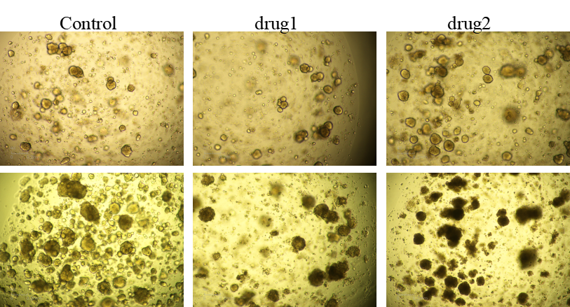
Conclusion: Human lung cancer organoids show general sensitivity to drug1; they are sensitive to drug2.
3. Organoid Drug Sensitivity - Human Pancreatic Cancer
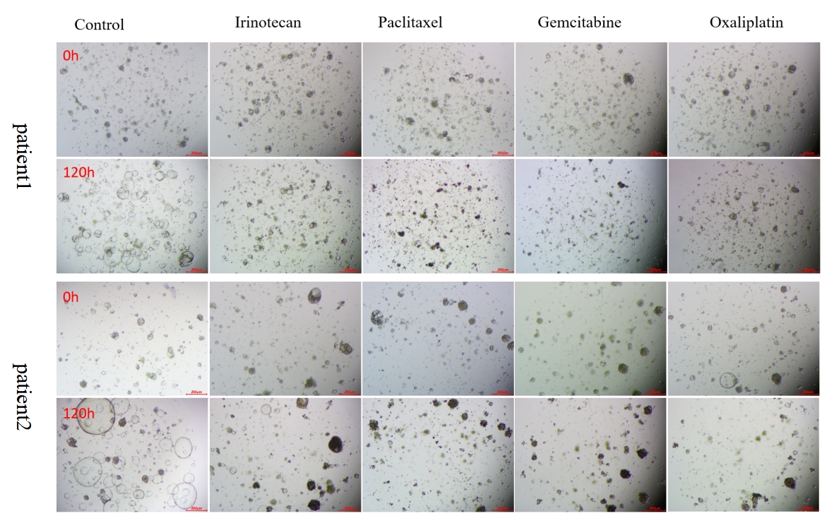
Conclusion: Human pancreatic cancer organoids show general sensitivity to Irinotecan and Oxaliplatin; they are sensitive to Paclitaxel and Gemcitabine; both patients exhibit consistent drug sensitivity effects.
4. Organoid Drug Sensitivity - Human Pancreatic Cancer

Conclusion: Human pancreatic cancer organoids show general sensitivity to Larotrectinib, Oclacitinib, Lenvatinib, and 5-Fluorouracil; both patients exhibit consistent drug sensitivity effects.
|
Item NO. |
Product Name |
Size |
|
Organoid ATP Viability Assay Kit |
100mL |
|
|
1kit |
||
|
1kit |
||
|
Mouse Lung Organoid Culture Medium Kit |
1kit |
|
|
Human Breast Cancer Organoid Culture Medium Kit |
1kit |
|
|
Human Endometrial carcinoma Organoid Culture Medium Kit |
1kit |
|
|
Human Colorectal cancer Organoid Culture Medium Kit |
1kit |
|
|
Human Lung Cancer Organoid Culture Medium Kit |
1kit |
|
|
1kit |
||
|
Human Pancreatic Cancer Organoid Culture Medium Kit |
1kit |
|
|
Human Ovarian Cancer Organoid Culture Medium Kit |
1kit |
|
|
Human Liver Organoid Culture Medium Kit |
1kit |
|
|
1kit |
||
|
Mouse Gastric Cancer Organoid Culture Medium Kit |
1kit |
|
|
1kit |
||
|
Mouse Breast Cancer Organoid Culture Medium Kit |
1kit |
|
|
Mouse Hepatocarcinoma Organoid Culture Medium Kit |
1kit |
|
|
1kit |
Absin provides antibodies, proteins, ELISA kits, cell culture, detection kits, and other research reagents. If you have any product needs, please contact us.
|
Absin Bioscience Inc. |
 Follow us on Facebook: Absin Bio Follow us on Facebook: Absin Bio |