|
Application of mIHC in exploring the characterization and function of different SUBtypes of T cells
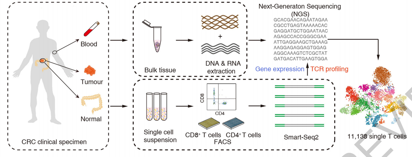
T cells are a key element in cancer immunotherapy, but certain basic properties, such as the development and migration of T cells within tumors, remain elusive. TCR is necessary for the recognition of foreign and autoantigens and can be used as a pedigree tag to track T cells in tumors. Here, we obtained transcripts from 11138 single T cells from 12 patients with colorectal cancer (CRC) and established the STARTRAC index (Single T cell analysis by RNA sequence and TCR tracking) to quantify the dynamic relationships among 20 functional and clonicity distinct T cell subsets.

In the literature, the authors used single-cell sequencing and flow cytometry to establish STARTRAC index, and used multiple fluorescence immunohistochemical techniques for location observation and in situ analysis.
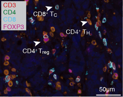
Multicolor IHC staining with CD3, CD4, CD8 and FOXP3 was used to verify the presence of T cells in colorectal cancer tumors
A representative example of colorectal cancer with multicolor IHC staining showed that anti-KI-67, CD8, PD-1 and Tim-3 were co-expressed in CD8+TEX cells

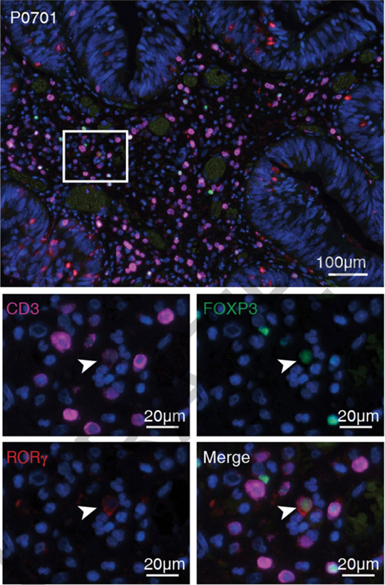
This is a typical example of a multicolor IHC staining of colorectal cancer. The white arrow shows the co-expression of CD3, FOXP3, and RORγ.
Multiple fluorescence immunohistochemistry literature interpretation ——summary
STARTRAC index analysis revealed various functional, migration, and developmental associations among different T cell subsets in colorectal cancer. The authors' data and previous findings in a mouse model reveal the TCR-dependent trajectory of tumor-infiltrating CD8+TEM cells on TEMRA and TEX, suggesting therapeutic strategies to promote the transformation of TEX to TEMRA.
The enrichment of CXCL13+BHLHE40+IFNG+ Th1-like cells in MSI patients not only provides a theoretical basis for the high response rate of these patients to checkpoint blockade, but also leads to the consideration of the treatment of these cells. A collated dataset of differentially expressed genes, such as IGFLR1, available through the interactive portal http://crc.cancer-pku.cn, can serve as a resource for further exploration of T cells, as well as the identification of novel regulatory mechanisms and therapeutic targets.
[Apis mIHC Services]
* Services we can provide:
Absin multiplex fluorescence immunohistochemistry service provides the whole process of pre-validation - staining - photographing - analysis.
Multi-color immunohistochemistry service with up to six indicators and seven colors (including DAPI) (chip with up to four indicators and five colors), samples include paraffin section, frozen section and tissue chip (TMA). For large tissue sections, please provide a single tissue section, FFPE sample, thickness 3-5um. For formal experiment, it is best to have 2 sheets for each case and 1 sheet for reserve. The number of pre-experimental samples is determined according to the pre-experimental scheme.
(* All antibody indicators need to be provided by you)
* Service Results we can provide:
1.5 colors and below provide 200X full image (equivalent to 20X objective lens and 10X eyepiece) original image, 6 or 7 colors provide 200X image with designated field of vision, generally 3 field of vision is default, beyond the field of vision will be charged separately, the charge standard is set according to the specific situation, the picture viewing software can be opened in the client. Data can be uploaded to an online disk with less than 1 gb, or sent to a USB flash disk with more than 1 GB.
2. Single-cell analysis results of the customer's designated area:
- Single positive cells occupy percentage of nuclear cells, density.
- Double positive cells occupy percentage of nuclear cells, density.
- Tissue area and specific pathological morphology area were calculated.
- TMA provides the above quantitative results for each core point.
Abs Multiplex fluorescent immunohistochemical products: four to seven color multicolor kits
Component information: TSA fluorescent dye, signal amplification reaction solution, pika generic or anti-rabbit HRP labeled secondary antibody, anti-fluorescence quenching sealing tablet, DAPI.
|
Absin Bioscience Inc.
TEL: +86-21-38015121
E-MAIL: worldwide@absin.cn
|
|